


Episode 14: Stargardt Disease — 유전성 황반변성의 원인과 ABCA4

Haewon Park
임상유전체학 연구원
최신 유전체 연구를 임상적 맥락에서 해석하고, 유전 데이터 속에 담긴 단서를 환자의 진단으로 연결하는 과정을 보다 쉽게 이해할 수 있도록 전달합니다.
시야의 중심이 점차 흐려지고, 읽기나 얼굴을 알아보는 것이 어려워진다면 망막의 중심부인 황반(macula)의 이상을 의심할 수 있습니다. 황반에 영향을 미치는 질환은 다양한데, 그중 Stargardt disease는 소아·청소년기에 시작되는 대표적인 유전성 황반변성(inherited macular degeneration)입니다.
이번 Clinical Series에서는 Stargardt disease의 주요 임상 특징과 함께, ABCA4 유전자 변이가 어떻게 망막에 lipofuscin을 축적시키고 시력 저하로 이어지는지 살펴보겠습니다.

자주 묻는 질문
Q. Stargardt disease는 어떻게 유전되나요?
A. 가장 대표적인 형태인 Stargardt disease 1(STGD1)은 상염색체 열성(autosomal recessive) 으로 유전되며, 일반적으로 양쪽 ABCA4 allele에 질환을 유발하는 변이가 존재할 때 발생합니다.
Q. 환자마다 발병 연령과 진행 속도가 다른 이유는 무엇인가요?
A. 주로 소아기 또는 청소년기에 증상이 시작되지만, 실제 발병 연령과 진행 속도는 환자마다 상당한 차이를 보입니다. ABCA4 변이의 종류와 잔존 기능 역시 이러한 임상적 다양성에 영향을 줄 수 있습니다.
Q. 초기에도 주변 시야가 함께 나빠지나요?
A. 질환 초기에는 주변부 망막이 상대적으로 보존되어 peripheral visual field가 정상으로 유지되는 경우가 많습니다. 먼저 영향을 받는 것은 망막 중심부의 황반입니다.
Q. 안저 및 영상 검사에서는 어떤 소견이 관찰되나요?
A. 안저검사에서는 황반 주변에 특징적인 yellow-white flecks가 관찰될 수 있으며, 물고기 모양을 닮았다고 표현되는 pisciform flecks는 Stargardt disease를 시사하는 대표적인 안저 소견 중 하나입니다.안저자가형광검사(fundus autofluorescence)에서는 lipofuscin 축적과 관련된 과자가형광(hyperautofluorescence) 반점과 망막색소상피(RPE) 위축 부위의 저자가형광(hypoautofluorescence)이 관찰될 수 있습니다. 이, OCT에서는 fovea의 thinning과 함께 photoreceptor 및 RPE layer의 위축이 관찰될 수 있습니다.
Q. Stargardt disease에서 유전자 검사는 어떤 도움이 되나요?
A. 임상적으로 유사한 다른 inherited retinal disease와의 감별뿐 아니라, ABCA4 변이의 확인과 유전 방식의 이해, 그리고 환자 및 가족에 대한 유전 상담에도 중요한 정보를 제공할 수 있습니다.
1. Stargardt disease란?
Stargardt disease는 주로 ABCA4 유전자의 병원성 변이에 의해 발생하는 유전성 망막질환입니다. 가장 대표적인 형태인 Stargardt disease 1(STGD1) 은 상염색체 열성(autosomal recessive)으로 유전되며, 일반적으로 양쪽 ABCA4 allele에 질환을 유발하는 변이가 존재할 때 발생합니다.
주로 소아기 또는 청소년기에 증상이 시작되지만, 실제 발병 연령과 질환의 진행 속도는 환자마다 상당한 차이를 보입니다.
변이의 종류와 잔존 기능이 표현형에 영향을 준다
ABCA4 변이의 종류와 잔존 기능 역시 이러한 임상적 다양성에 영향을 줄 수 있습니다.

2. 중심 시력부터 영향을 받는 Stargardt disease
Stargardt disease의 대표적인 특징은 진행성 중심 시력 저하(progressive central vision loss) 입니다.
망막 중심부의 황반, 특히 photoreceptor와 retinal pigment epithelium(RPE) 이 점차 손상되면서 세밀한 물체를 구분하거나 글씨를 읽고 얼굴을 인식하는 능력이 저하될 수 있습니다.
반면 질환 초기에는 주변부 망막이 상대적으로 보존되어 peripheral visual field가 정상으로 유지되는 경우가 많습니다.
- 검사에서 관찰되는 소견
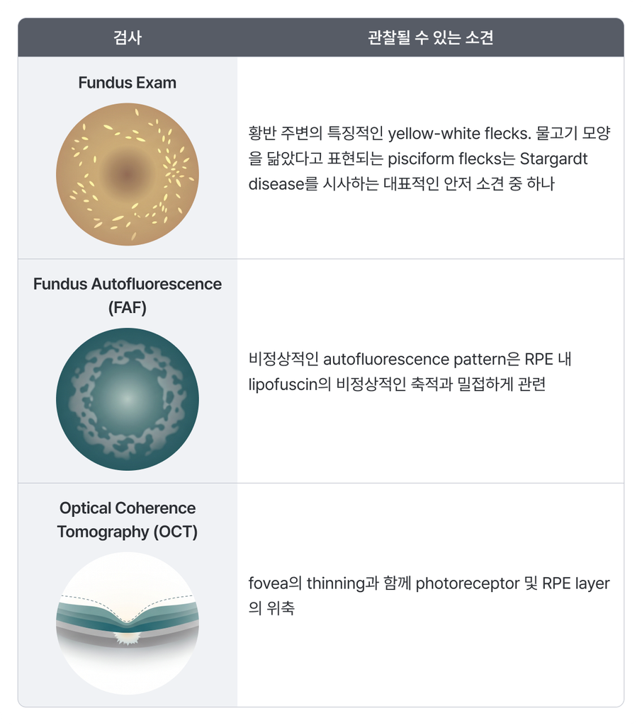
그렇다면 ABCA4의 이상은 어떻게 이러한 특징적인 망막 손상을 만들어낼까요?
3. ABCA4: photoreceptor의 중요한 transporter
ABCA4는 약 2,273개의 amino acid로 이루어진 대형 ATP-binding cassette transporter입니다. ABCA4 단백질은 photoreceptor outer segment의 disc membrane에 위치하며, 여러 기능적 domain을 통해 정상적인 visual cycle을 유지하는 데 중요한 역할을 합니다.
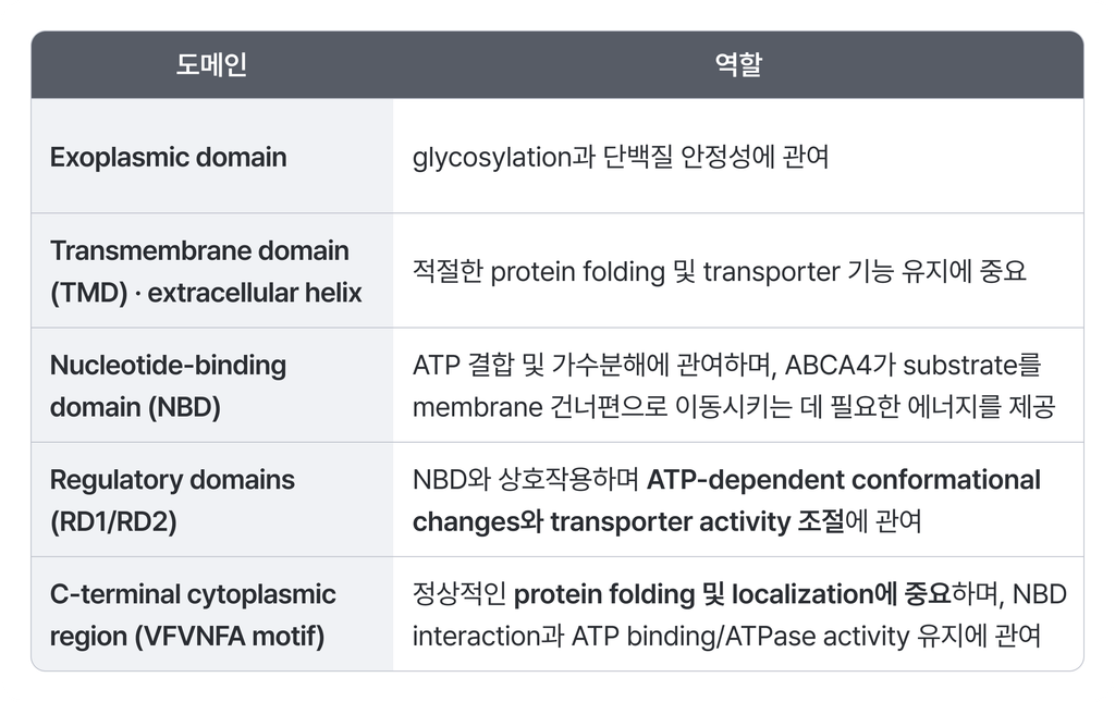
따라서 ABCA4의 서로 다른 영역에서 발생한 변이는 protein folding, expression, ATPase activity 또는 substrate transport 등에 다양한 영향을 미칠 수 있으며, 결과적으로 환자별 표현형의 차이로 이어질 수 있습니다.
4. 시각을 유지하는 Visual Cycle
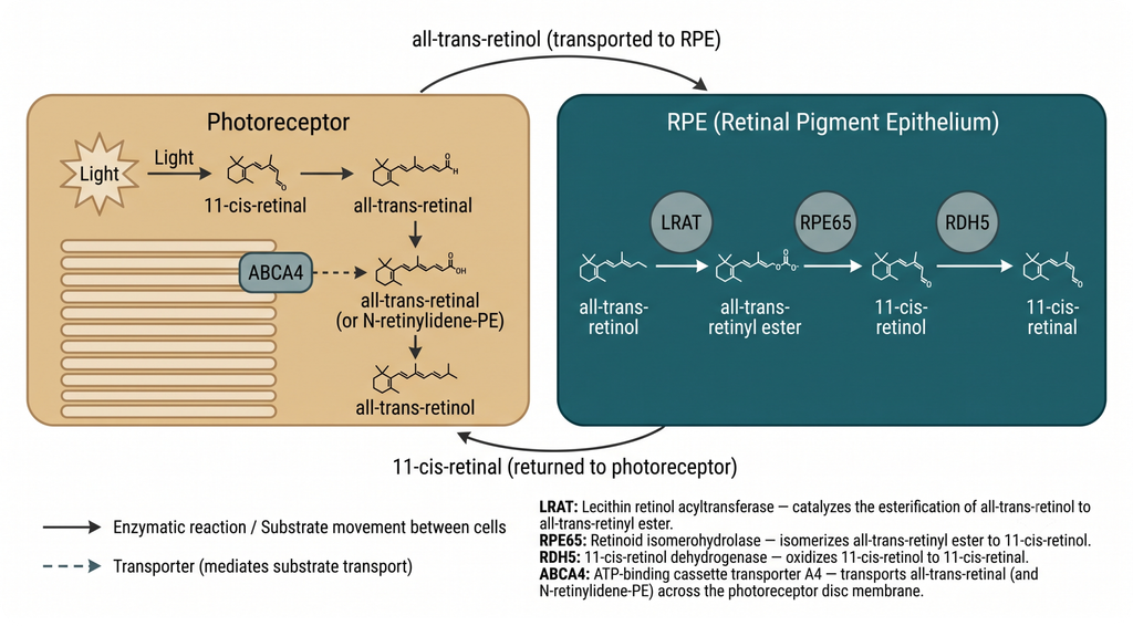
빛을 감지한 photoreceptor에서는 11-cis-retinal이 all-trans-retinal로 전환됩니다. 이 과정은 우리가 빛을 인지하는 phototransduction의 핵심이지만, 생성된 all-trans-retinal은 다시 처리되어야 합니다.
Photoreceptor에서 생성된 retinoid는 RPE와 photoreceptor 사이의 visual cycle을 통해 재활용됩니다. RPE에서는 LRAT, RPE65, RDH5 등의 단백질이 관여하여 retinoid를 다시 11-cis-retinal 형태로 재생하고, 이는 photoreceptor에서 새로운 photopigment를 형성하는 데 사용됩니다.
이 과정에서 ABCA4는 photoreceptor disc에 남아 있는 all-trans-retinal의 제거를 돕는 중요한 역할을 담당합니다.
5. ABCA4가 제대로 작동하지 않는다면?
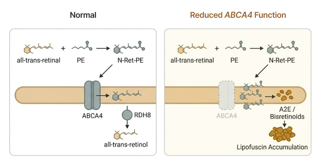
a. 정상적인 경우
Photoreceptor disc 내부에서 all-trans-retinal은 phosphatidylethanolamine(PE) 과 결합하여 N-retinylidene-phosphatidylethanolamine(N-Ret-PE) 을 형성할 수 있습니다.
정상적인 ABCA4는 ATP를 이용하여 N-Ret-PE를 disc membrane의 lumenal leaflet에서 cytoplasmic leaflet으로 이동시키는 데 관여합니다. 이후 N-Ret-PE가 all-trans-retinal과 PE로 해리되면, all-trans-retinal은 RDH8 등의 효소에 의해 all-trans-retinol로 환원되어 정상적인 retinoid cycle로 다시 들어갈 수 있습니다.
b. 기능이 감소하면
ABCA4의 기능이 감소하면 N-Ret-PE와 retinal-derived compounds가 충분히 제거되지 못합니다. 축적된 retinal derivatives는 여러 반응을 거쳐 A2E와 같은 bisretinoid compounds를 형성하고, 이들이 RPE에 축적되면서 lipofuscin의 주요 구성 성분이 됩니다.
Stargardt disease 환자의 RPE에서는 정상보다 현저하게 많은 lipofuscin이 축적될 수 있습니다.
6. Lipofuscin 축적에서 시력 저하까지
A2E 등의 bisretinoid를 포함하는 lipofuscin의 과도한 축적은 단순히 세포 내부에 노폐물이 쌓이는 것에서 끝나지 않습니다. 이러한 물질의 축적은 RPE의 정상적인 기능을 방해하고 oxidative stress와 세포독성을 증가시킬 수 있습니다. 시간이 지나면서 RPE가 손상되고, RPE의 기능에 의존하는 photoreceptor 역시 점차 소실될 수 있습니다.
결국 다음과 같은 일련의 과정이 Stargardt disease의 핵심 병태생리를 설명합니다.
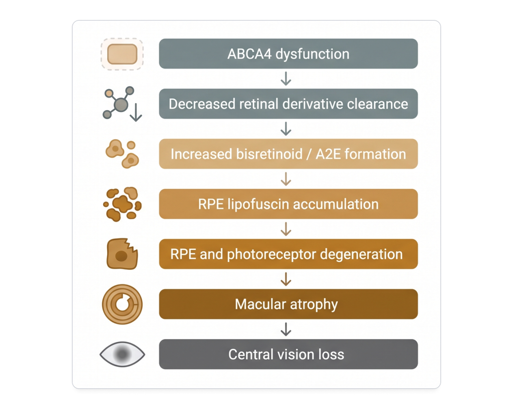
안저에서 관찰되는 특징적인 yellow-white flecks 역시 이러한 비정상적인 lipofuscin 및 bisretinoid 축적과 관련되어 있습니다.
7. 유전자 변이가 설명하는 Stargardt disease
Stargardt disease는 하나의 유전자 이상이 어떻게 특정 조직의 대사 과정에 영향을 주고, 결국 특징적인 임상 표현형으로 이어지는지를 잘 보여주는 유전질환입니다.
ABCA4는 photoreceptor에서 visual cycle 중 발생하는 retinal derivatives를 처리하는 데 중요한 역할을 합니다. 이 기능이 저하되면 bisretinoid와 lipofuscin이 RPE에 비정상적으로 축적되고, 장기적으로 photoreceptor와 RPE의 degeneration을 유발합니다.
유전자 검사가 제공하는 정보
따라서 Stargardt disease에서 유전자 검사는 다음과 같은 정보를 제공할 수 있습니다.
- 임상적으로 유사한 다른 inherited retinal disease와의 감별
- Stargardt disease의 원인이 되는 ABCA4 변이의 확인
- 가족검사를 통한 변이의 phase 및 유전 양상 확인
- 환자 및 가족에 대한 유전 상담
마무리
Stargardt disease의 특징적인 중심 시력 저하와 황반의 변화 뒤에는 결국 photoreceptor의 작은 transporter 하나가 정상적인 visual cycle을 유지하는 정교한 과정이 숨어 있습니다.
이것이 Stargardt disease를 이해할 때 ABCA4와 visual cycle을 함께 살펴봐야 하는 이유입니다.
희귀질환 변이 해석, 어떻게 하고 계신가요?
GEBRA의 판독 환경을 무료로 직접 확인해 보실 수 있습니다.
참고 문헌
Abalem MF, Otte B, Andrews C, et al. Peripheral visual fields in ABCA4 Stargardt disease and correlation with disease extent on ultra-widefield fundus autofluorescence. Am J Ophthalmol. 2017.
Strauss RW, Kong X, Ho A, et al. Progression of Stargardt disease as determined by fundus autofluorescence over a 12-month period: ProgStar Report No. 11. JAMA Ophthalmol. 2019.
Heath Jeffery RC, Chen FK. Stargardt disease: Multimodal imaging: A review. Clin Exp Ophthalmol. 2021.
Curtis SB, Molday LL, Garces FA, Molday RS. Functional analysis and classification of homozygous and hypomorphic ABCA4 variants associated with Stargardt macular degeneration. Hum Mutat. 2020.
Al-Khuzaei S, Broadgate S, Foster CR, et al. An overview of the genetics of ABCA4 retinopathies, an evolving story. Genes (Basel). 2021.
Zhong M, Molday LL, Molday RS. Role of the C terminus of the photoreceptor ABCA4 transporter in protein folding, function, and retinal degenerative diseases. J Biol Chem. 2009.
Scortecci JF, Garces FA, Mahto JK, Molday LL, Van Petegem F, Molday RS. Structural and functional characterization of the nucleotide-binding domains of ABCA4 and their role in Stargardt disease. J Biol Chem. 2024.
Tripathy K, Kaur K. Stargardt Disease. StatPearls. 2024.
Molday RS, Garces FA, Scortecci JF, Molday LL. Structure and function of ABCA4 and its role in the visual cycle and Stargardt macular degeneration. Prog Retin Eye Res. 2022.
Giacalone JC, Scruggs BA. The pathology of Stargardt disease and future treatment horizons. Retinal Physician. 2024.
이 글의 시리즈
더보기