



Episode 15: Homocystinuria — 호모시스틴뇨증의 원인 유전자와 감별 포인트

Medical Genetics Group
3billion
환자 유전체 분석을 통해 희귀질환 원인 변이를 발굴하고 임상적 해석 및 연구 역량 강화에 기여합니다.
우리 몸에서는 음식으로 섭취한 methionine이 여러 대사 과정을 거치며 사용됩니다. 이 과정에서 생성되는 중요한 중간 대사산물 중 하나가 homocysteine입니다.
정상적인 상황에서 homocysteine은 다시 methionine으로 전환되는 remethylation pathway 또는 cystathionine을 거쳐 cysteine으로 전환되는 transsulfuration pathway를 통해 대사됩니다.
그러나 이러한 대사 과정에 관여하는 효소나 보조인자의 기능에 문제가 발생하면 homocysteine이 정상적으로 처리되지 못하고 체내에 축적될 수 있습니다. 이처럼 혈액 내 homocysteine 농도가 크게 증가하고 소변에서도 homocysteine/homocystine이 증가하는 유전성 대사질환들을 호모시스틴뇨증(homocystinuria) 이라고 합니다.
자주 묻는 질문
Q. Homocysteine이 증가하면 항상 CBS deficiency인가요?
A. 아닙니다. Homocysteine을 다시 methionine으로 전환하는 remethylation pathway에 문제가 발생해도 호모시스틴뇨증이 나타날 수 있습니다. 대표적으로 MTHFR deficiency에서는 5-methyltetrahydrofolate 생성이 감소하면서 remethylation이 저하됩니다.
Q. CBS deficiency와 remethylation 장애는 어떻게 구분하나요?
methionine이 중요한 단서입니다. CBS deficiency에서는 transsulfuration이 저하되면서 homocysteine과 methionine이 함께 증가하는 것이 특징적입니다. 반면 MTHFR, MTR, MTRR 및 cobalamin metabolism과 관련된 질환에서는 remethylation이 저하되므로 homocysteine은 증가하지만 methionine은 낮거나 정상 범위일 수 있습니다.
Q. 왜 methylmalonic acid까지 함께 확인해야 하나요?
A. MMACHC를 비롯해 adenosylcobalamin과 methylcobalamin pathway를 동시에 침범하는 질환에서는 homocysteine 상승에 더해 methylmalonic acid elevation까지 동반될 수 있기 때문입니다.
Q. 대사효소를 직접 코딩하는 유전자가 아니어도 원인이 될 수 있나요?
A. 될 수 있습니다. 예를 들어 HCFC1은 MMACHC expression 조절과 연관되어 있으며, pathogenic variants는 X-linked cobalamin disorder인 Methylmalonic aciduria and homocystinemia, cblX type를 일으킬 수 있습니다. 또한 특정 PRDX1 영역의 변이에 의해 비정상적인 read-through transcription이 발생하면 인접한 MMACHC의 promoter methylation과 transcriptional silencing을 유발할 수 있습니다.
1. Homocysteine은 어떻게 대사될까요?
Homocysteine 대사는 크게 두 방향으로 이루어집니다.
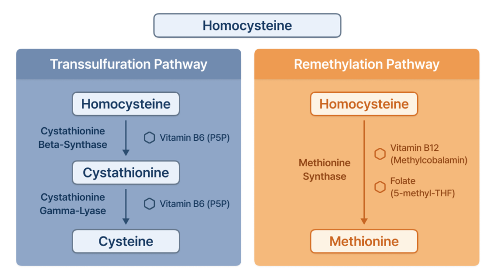
따라서 CBS, MTHFR, MTR, MTRR과 같은 유전자뿐만 아니라 vitamin B12의 세포 내 processing 및 trafficking에 관여하는 유전자에 이상이 발생해도 심한 hyperhomocysteinemia 또는 호모시스틴뇨증이 나타날 수 있습니다.
2. Homocysteine이 축적되면 어떤 문제가 생길까요?
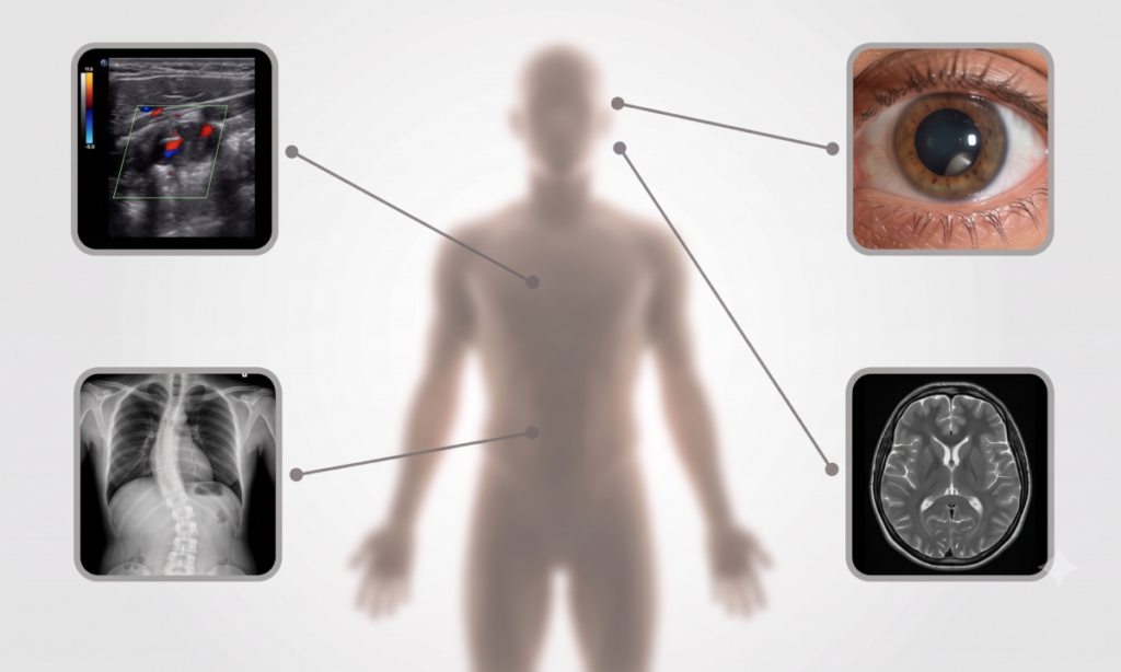
Homocysteine의 과도한 축적은 여러 조직에 영향을 줄 수 있습니다.
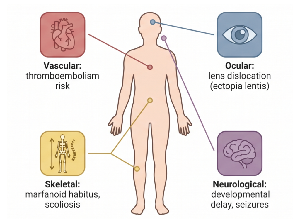
환자마다 증상의 조합과 중증도는 크게 다릅니다. 따라서 호모시스틴뇨증은 단순히 하나의 장기에 국한된 질환이라기보다 vascular, ocular, skeletal, and neurological systems에 걸쳐 다양한 증상을 일으킬 수 있는 multisystem metabolic disorder로 이해할 수 있습니다.
3. 대표적인 원인: CBS deficiency
가장 잘 알려진 형태는 CBS 유전자의 biallelic pathogenic variants로 발생하는 cystathionine beta-synthase deficiency입니다.
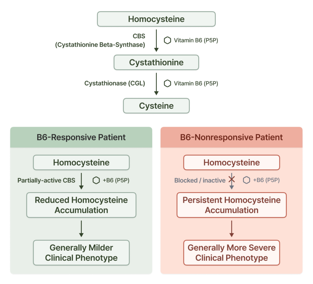
CBS 기능이 감소하면 homocysteine이 cystathionine으로 정상적으로 전환되지 못합니다. 그 결과 plasma total homocysteine이 크게 증가하고, 일반적으로 methionine 역시 증가합니다.
CBS deficiency는 autosomal recessive disorder이며, 같은 질환에서도 임상 양상은 매우 다양합니다.
3-1. Pyridoxine responsiveness
특히 중요한 특징 중 하나는 pyridoxine(vitamin B6) responsiveness입니다.
일부 환자에서는 pyridoxine 투여에 의해 CBS의 residual activity가 증가하면서 homocysteine 농도가 크게 감소합니다. 이러한 B6-responsive 환자는 일반적으로 B6-nonresponsive 환자보다 임상 양상이 경한 경향이 있지만, 실제 responsiveness는 genotype만으로 단정하기보다 실제 response를 통해 평가하는 것이 중요합니다.
따라서 CBS deficiency가 의심되는 환자에서는 biochemical findings와 molecular genetic findings뿐만 아니라 pyridoxine response를 함께 평가하는 것이 중요합니다.
4. 모든 호모시스틴뇨증이 CBS deficiency인 것은 아닙니다
Homocysteine이 증가했다고 해서 항상 CBS에 문제가 있는 것은 아닙니다. Homocysteine을 다시 methionine으로 전환하는 remethylation pathway에 문제가 발생해도 호모시스틴뇨증이 나타날 수 있습니다.
대표적으로 MTHFR deficiency에서는 5-methyltetrahydrofolate 생성이 감소하면서 remethylation이 저하됩니다. 이 경우 severe MTHFR deficiency에서는 homocysteine이 크게 증가하는 반면, methionine은 일반적으로 감소합니다.
“이는 CBS deficiency에서 homocysteine과 methionine이 모두 증가하는 것과 대비되는 중요한 biochemical clue입니다.”
MTR과 MTRR의 pathogenic variants 역시 methionine synthase system을 방해하여 호모시스틴뇨증을 일으킬 수 있습니다. 이러한 질환에서도 impaired remethylation으로 인해 hyperhomocysteinemia와 low or normal methionine이 나타날 수 있습니다.
5. Cobalamin metabolism과 관련된 원인들
Homocysteine metabolism에는 vitamin B12가 중요한 역할을 합니다. 세포 내로 들어온 cobalamin은 여러 단계를 거쳐 활성 cofactor로 처리되어야 합니다.
이 과정에 문제가 발생하면 methionine synthase에 필요한 methylcobalamin을 제대로 공급하지 못하면서 homocysteine의 remethylation이 저하될 수 있습니다.
5-1. MMACHC와 Methylmalonic aciduria and homocystinuria, cblC type
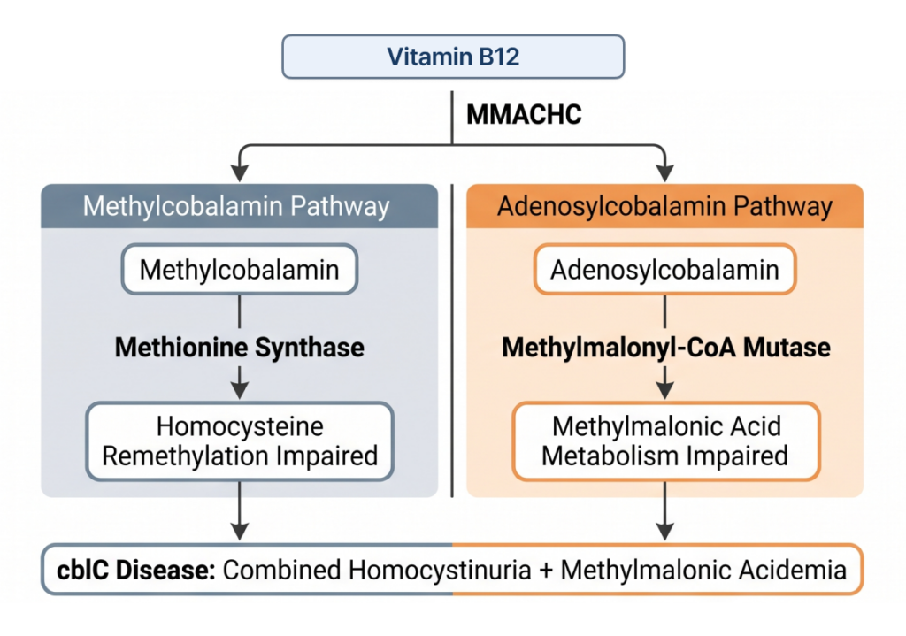
대표적인 유전자가 MMACHC입니다. MMACHC-related Methylmalonic aciduria and homocystinuria, cblC type에서는 methylcobalamin뿐 아니라 adenosylcobalamin metabolism도 영향을 받을 수 있기 때문에 호모시스틴뇨증과 methylmalonic acidemia가 함께 나타나는 것이 특징적입니다.
환자에서는 developmental delay, neurologic abnormalities, hematologic abnormalities, ophthalmologic manifestations 및 여러 장기의 이상이 나타날 수 있으며, infantile-onset부터 late-onset까지 임상 양상이 다양합니다.
5-2. 같은 MMADHC 유전자라도 변이 위치에 따라 phenotype이 달라집니다
MMADHC는 cobalamin metabolism에서 특히 흥미로운 유전자입니다. MMADHC pathogenic variants는 변이의 위치와 기능적 영향에 따라 서로 다른 biochemical phenotype을 나타낼 수 있습니다.
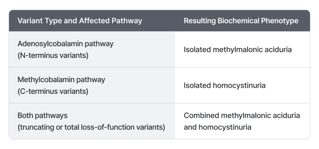
따라서 MMADHC에서는 단순히 “어느 유전자에 변이가 있는가” 뿐만 아니라 “변이가 단백질의 어느 부분에 위치하고 어떤 기능을 손상시키는가” 가 phenotype을 이해하는 데 중요합니다.
5-3. 직접적인 대사효소가 아닌 유전자도 원인이 될 수 있습니다
호모시스틴뇨증과 combined methylmalonic acidemia/homocystinuria의 원인이 반드시 homocysteine을 직접 catalyze하는 효소에 국한되는 것은 아닙니다.
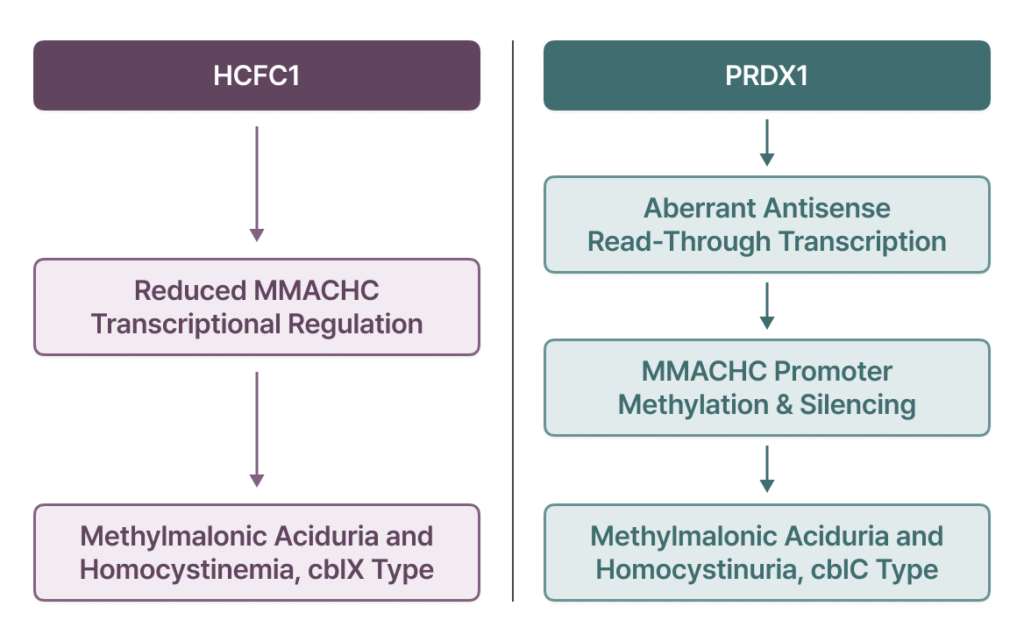
즉, metabolic disease에서는 특정 효소의 coding sequence뿐만 아니라 gene expression과 epigenetic regulation을 변화시키는 유전적 이상 역시 질환의 원인이 될 수 있습니다.
6. 원인 pathway는 어떻게 감별할까요?
호모시스틴뇨증에서 중요한 점은 같은 “homocysteine elevation” 이라는 biochemical phenotype을 보여도 underlying mechanism이 서로 다를 수 있다는 것입니다.
- CBS deficiency — transsulfuration이 저하되면서 homocysteine과 methionine이 함께 증가하는 것이 특징적입니다.
- MTHFR, MTR, MTRR 및 methylcobalamin metabolism 관련 질환 — remethylation이 저하되므로 homocysteine은 증가하지만 methionine은 낮거나 정상 범위일 수 있습니다.
- MMACHC를 비롯해 adenosylcobalamin과 methylcobalamin pathway를 동시에 침범하는 질환 — 여기에 methylmalonic acid elevation까지 동반될 수 있습니다.
따라서 plasma total homocysteine만 확인하는 것보다 methionine과 methylmalonic acid를 함께 평가하는 것이 원인 감별에 매우 중요합니다.
6-1. Biochemical profile로 원인 pathway 추정하기
심한 hyperhomocysteinemia가 확인되었다면 biochemical profile을 바탕으로 원인이 되는 pathway를 추정할 수 있습니다.
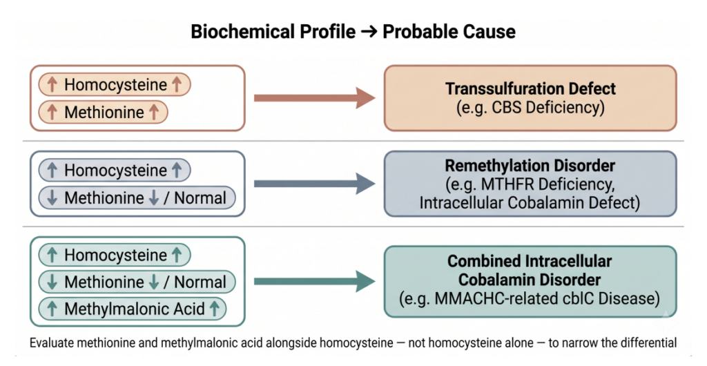
이러한 biochemical findings에 환자의 임상 증상과 genetic testing 결과를 함께 해석하면 보다 정확한 진단에 접근할 수 있습니다.
Take-home message
호모시스틴뇨증은 하나의 단일 질환이라기보다 homocysteine metabolism에 영향을 미치는 여러 유전질환에서 나타날 수 있는 중요한 biochemical phenotype입니다.
가장 대표적인 원인은 CBS deficiency이지만, MTHFR, MTR, MTRR과 같은 remethylation pathway의 유전자뿐만 아니라 MMACHC, MMADHC, HCFC1 등 intracellular cobalamin metabolism과 관련된 다양한 유전자 역시 원인이 될 수 있습니다.
또한 같은 pathway 또는 같은 유전자에 이상이 있더라도 변이의 위치와 residual function에 따라 onset과 severity, biochemical profile이 달라질 수 있습니다.
따라서 호모시스틴뇨증이 의심되는 환자에서는 homocysteine의 증가 자체뿐만 아니라 methionine과 methylmalonic acid의 변화, 임상 phenotype, 그리고 유전적 원인을 종합적으로 평가하는 것이 중요합니다.
이러한 접근은 복잡한 homocysteine metabolism disorder에서 정확한 원인을 파악하고 적절한 진단과 치료 방향을 결정하는 데 중요한 단서가 될 수 있습니다.
GEBRA는 이러한 희귀질환 변이 해석 과정을 지원하는 플랫폼으로, 무료로 직접 확인해 보실 수 있습니다.
희귀질환 변이 해석, 어떻게 하고 계신가요?
GEBRA의 판독 환경을 무료로 직접 확인해 보실 수 있습니다.
참고문헌
Althubity, A. A. (2025). Homocystinuria: Advances in metabolic and molecular therapies targeting homocysteine pathways. Molecular Medicine Reports, 33(1), 34.
Esse, R., Barroso, M., Tavares de Almeida, I., & Castro, R. (2019). The contribution of homocysteine metabolism disruption to endothelial dysfunction: State-of-the-art. International Journal of Molecular Sciences, 20(4), 867.
Frosst, P., Blom, H. J., Milos, R., Goyette, P., Sheppard, C. A., Matthews, R. G., et al. (1995). A candidate genetic risk factor for vascular disease: A common mutation in methylenetetrahydrofolate reductase. Nature Genetics, 10(1), 111–113.
Goyette, P., Sumner, J. S., Milos, R., Duncan, A. M., Rosenblatt, D. S., Matthews, R. G., & Rozen, R. (1994). Human methylenetetrahydrofolate reductase: Isolation of cDNA, mapping and mutation identification. Nature Genetics, 7(2), 195–200.
Guéant, J. L., Chéry, C., Oussalah, A., Nadaf, J., Coelho, D., Josse, T., et al. (2018). A PRDX1 mutant allele causes a MMACHC secondary epimutation in cblC patients. Nature Communications, 9(1), 67.
Gupta, S., Kelow, S., Wang, L., Andrake, M. D., Dunbrack, R. L., & Kruger, W. D. (2018). Mouse modeling and structural analysis of the p.G307S mutation in human cystathionine β-synthase (CBS) reveal effects on CBS activity but not stability. Journal of Biological Chemistry, 293(36), 13921–13931.
Jakubowski, H., Zhang, L., Bardeguez, A., & Aviv, A. (2000). Homocysteine thiolactone and protein homocysteinylation in human endothelial cells: Implications for atherosclerosis. Circulation Research, 87(1), 45–51.
Koklesova, L., Mazurakova, A., Samec, M., Biringer, K., Samuel, S. M., Büsselberg, D., et al. (2021). Homocysteine metabolism as the target for predictive medical approach, disease prevention, prognosis, and treatments tailored to the person. EPMA Journal, 12(4), 477–505.
Kraus, J. P., Janošík, M., Kožich, V., Mandell, R., Shih, V., Sperandeo, M. P., et al. (1999). Cystathionine β-synthase mutations in homocystinuria. Human Mutation, 13(5), 362–375.
Lerner-Ellis, J. P., Anastasio, N., Liu, J., Coelho, D., Suormala, T., Stucki, M., et al. (2009). Spectrum of mutations in MMACHC, allelic expression, and evidence for genotype–phenotype correlations. Human Mutation, 30(7), 1072–1081.
Lerner-Ellis, J. P., Tirone, J. C., Pawelek, P. D., Doré, C., Atkinson, J. L., Watkins, D., et al. (2006). Identification of the gene responsible for methylmalonic aciduria and homocystinuria, cblC type. Nature Genetics, 38(1), 93–100.
Poddar, R., & Paul, S. (2009). Homocysteine–NMDA receptor-mediated activation of extracellular signal-regulated kinase leads to neuronal cell death. Journal of Neurochemistry, 110(3), 1095–1106.
Pizzorno, J. (2014). Homocysteine: Friend or foe? Integrative Medicine: A Clinician’s Journal, 13(4), 8.
Ruiz-Mercado, M., Vargas, M. T., de Soto, I. P., Pecellín, C. D., Sánchez, M. C., Delgado, M. B., et al. (2016). Methionine synthase reductase deficiency (CblE): A report of two patients and a novel mutation. Hematology, 21(3), 193–197.
Sacharow, S. J., & Levy, H. L. (2025). Homocystinuria due to cystathionine beta-synthase deficiency. In GeneReviews®.
Sauls, D. L., Wolberg, A. S., & Hoffman, M. (2003). Elevated plasma homocysteine leads to alterations in fibrin clot structure and stability: Implications for the mechanism of thrombosis in hyperhomocysteinemia. Journal of Thrombosis and Haemostasis, 1(2), 300–306.
Stucki, M., Coelho, D., Suormala, T., Burda, P., Fowler, B., & Baumgartner, M. R. (2012). Molecular mechanisms leading to three different phenotypes in the cblD defect of intracellular cobalamin metabolism. Human Molecular Genetics, 21(6), 1410–1418.
Torices, L., de Las Heras, J., Arango-Lasprilla, J. C., Cortés, J. M., Nunes-Xavier, C. E., & Pulido, R. (2021). MMADHC premature termination codons in the pathogenesis of cobalamin D disorder: Potential of translational readthrough reconstitution. Molecular Genetics and Metabolism Reports, 26, 100710.
Watkins, D., Ru, M., Hwang, H. Y., Kim, C. D., Murray, A., Philip, N. S., et al. (2002). Hyperhomocysteinemia due to methionine synthase deficiency, cblG: Structure of the MTR gene, genotype diversity, and recognition of a common mutation, P1173L. The American Journal of Human Genetics, 71(1), 143–153.
Yu, H. C., Sloan, J. L., Scharer, G., Brebner, A., Quintana, A. M., Achilly, N. P., et al. (2013). An X-linked cobalamin disorder caused by mutations in transcriptional coregulator HCFC1. The American Journal of Human Genetics, 93(3), 506–514.
이 글의 시리즈
더보기