Episode 16: Maple Syrup Urine Disease (MSUD): Causative Genes and Branched-Chain Amino Acid Metabolism

Medical Genetics Group
3billion
We contribute to the discovery of causative variants in rare diseases through patient genome analysis, while strengthening clinical interpretation and research capabilities.
What condition would you suspect if a newborn’s urine or earwax carried a sweet smell resembling maple syrup?
Maple syrup urine disease (MSUD) is a rare autosomal recessive disorder caused by a defect in the metabolism of branched-chain amino acids (BCAAs). Leucine, isoleucine and valine are not broken down normally, so these amino acids and their metabolites accumulate in the body, and in severe cases the condition can progress to life-threatening metabolic encephalopathy.
MSUD was first described in 1954, when John H. Menkes and colleagues reported four affected infants in a single family. The patients showed progressive neurological deterioration together with the characteristic sweet-smelling urine, and the disease name comes from that feature.
The incidence in the general population is reported as approximately 1 in 185,000. Clinical severity, however, varies widely between patients, ranging from severe neurological symptoms immediately after birth to relatively mild symptoms appearing during growth.
These differences are influenced by several factors, including the causative gene and the nature of the variant, residual enzyme activity, and the state of metabolic control.

FAQs
Q. Which genes cause maple syrup urine disease?
A. MSUD is caused mainly by biallelic pathogenic variants in BCKDHA, BCKDHB and DBT. All three encode component proteins of the mitochondrial BCKD complex.
Q. Can MSUD be excluded if plasma BCAA levels are normal?
A. In intermittent MSUD, BCAA levels can be normal or near normal during metabolically stable periods. Even when biochemical results are unremarkable, if clinical suspicion persists it is important to interpret the metabolic state at the time of testing together with the genetic testing results.
Q. Why does alloisoleucine matter?
A. Its plasma concentration is very low under normal conditions, but it can rise in patients with MSUD, and it is a more disease-specific finding than a rise in BCAAs alone.
Q. Can severity be predicted from the causative gene?
A. Associations between causative gene and clinical severity have been reported, but they are not absolute. Variants in the same gene can produce a range of clinical phenotypes.
Q. What should be considered when only one pathogenic variant is identified?
A. In an autosomal recessive disorder, when only one pathogenic variant is found, it is necessary to consider a CNV on the other allele or a variant that is difficult to detect by standard sequence analysis.
1. Key Clinical Features of MSUD
The hallmark of MSUD is neurological impairment caused by the accumulation of branched-chain amino acids.
In classic MSUD, the most severe form, metabolic derangement progresses from immediately after birth, and without appropriate treatment severe neurological damage can occur within the first one to two weeks of life.
Early signs are non-specific, including poor feeding, irritability and lethargy. As metabolites continue to accumulate, abnormal muscle tone, opisthotonus and apnea can develop.
Some patients show characteristic abnormal movements such as bicycling movements or fencing posture.
With further progression, the condition can lead to cerebral edema, reduced consciousness, coma and central respiratory failure.
1-1. Clinical subtypes of MSUD
MSUD is divided into the following phenotypes by clinical severity.
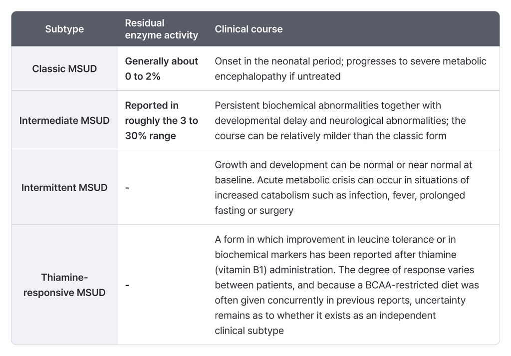
Patients with intermittent MSUD can have near-normal plasma amino acid levels at baseline, which may delay diagnosis.
Rather than being defined by a single clinical presentation, MSUD can be understood as a disease spectrum whose severity varies according to residual enzyme activity and the response to metabolic stress.
2. Causative Genes and Inheritance
MSUD is an autosomal recessive (AR) disorder caused mainly by biallelic pathogenic variants in BCKDHA, BCKDHB and DBT.
All of these genes encode component proteins of the mitochondrial branched-chain α-ketoacid dehydrogenase (BCKD) complex.
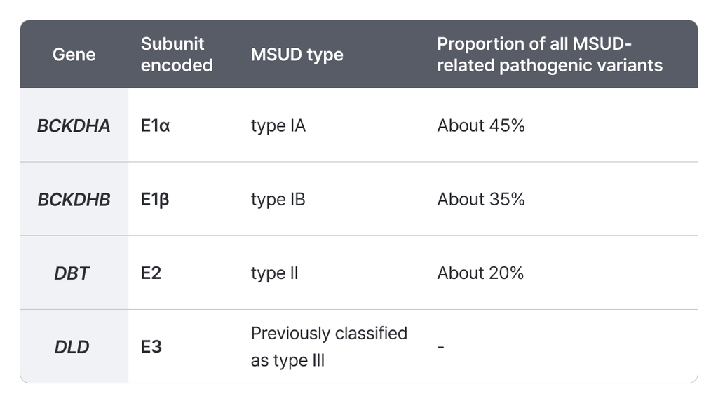
The E3 protein is shared across several mitochondrial enzyme complexes and not only the BCKD complex. DLD-related disease was previously classified as MSUD type III, but DLD deficiency is now generally treated as a separate metabolic disorder, distinct from classic MSUD.
3. Molecular Mechanism of MSUD: BCAA Metabolism and the BCKD Complex
Understanding the mechanism of MSUD starts with the normal metabolism of branched-chain amino acids.
Leucine, isoleucine and valine are essential amino acids that the body does not synthesize and that are supplied through food.
3-1. Normal BCAA pathway
- Leucine, isoleucine, valine → transamination → the corresponding branched-chain α-ketoacids (BCKAs)
- BCKAs → oxidative decarboxylation by the mitochondrial BCKD complex
- Products feed into acetyl-CoA, acetoacetate and succinyl-CoA, which are used in energy metabolism
The BCKD complex is a multienzyme complex made up of the components E1, E2 and E3.
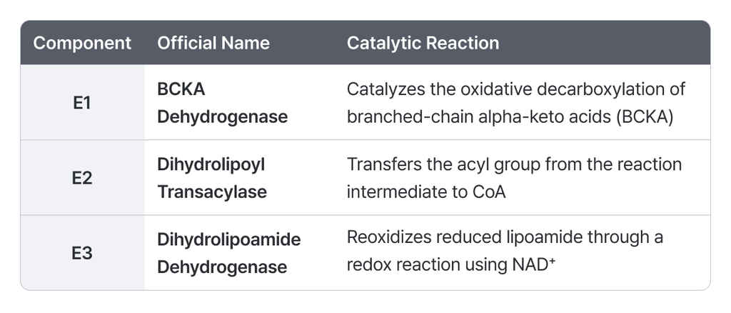
* The formal name of E1 is BCKA dehydrogenase. Because E1 carries out decarboxylation while the complex as a whole acts as a dehydrogenase, many sources write E1 as BCKA decarboxylase to distinguish the two. The naming may therefore differ from source to source.
3-2. What changes in MSUD
- Loss-of-function variants in BCKDHA, BCKDHB or DBT → reduced BCKD complex activity
- BCAAs and BCKAs accumulate without being broken down normally
- Rising plasma leucine → disruption of cerebral amino acid homeostasis, with effects on neurotransmitter synthesis and energy metabolism
- Cerebral edema and metabolic encephalopathy → coma or respiratory failure in severe cases
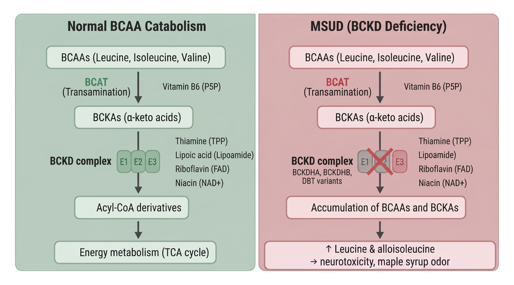
Excessive accumulation of leucine in particular is the central factor in the neurological damage seen in MSUD.
The pathophysiology of MSUD therefore has to be understood not as a simple rise in amino acid concentration, but as a combined process of mitochondrial metabolic failure and neurotoxicity.
4. Biochemical Diagnosis of MSUD and the Role of Alloisoleucine
In diagnosing MSUD, an increase in plasma branched-chain amino acids is an important clue.
Leucine, isoleucine and valine are generally elevated, and alloisoleucine in particular is an important biochemical marker for the diagnosis.
Alloisoleucine is a stereoisomer related to isoleucine metabolism, and its plasma concentration is very low under normal conditions. In patients with MSUD, by contrast, alloisoleucine can rise as a result of disordered BCAA metabolism, and this is a finding with higher disease specificity than a rise in BCAAs alone.
4-1. The usual diagnostic pathway for MSUD
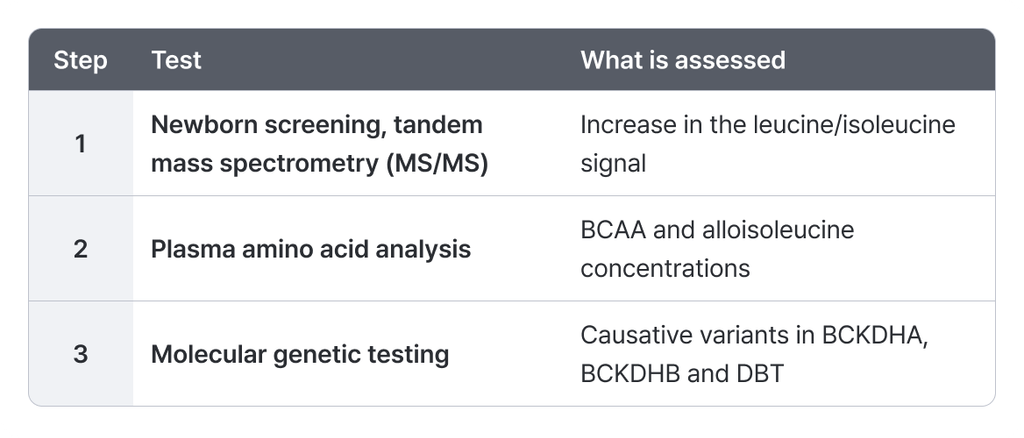
In intermittent MSUD, however, BCAA levels can be normal or near normal during metabolically stable periods.
Even when biochemical results are unremarkable, therefore, if clinical suspicion persists it is important to interpret the metabolic state at the time of testing together with the genetic testing results.
5. Pathogenic Variant Types in MSUD and Molecular Diagnosis
A range of pathogenic variant types has been reported in the MSUD causative genes.
These include single-nucleotide and small insertion/deletion variants such as missense, nonsense, frameshift and splice-altering variants. In some patients, copy number variants (CNVs) such as exon-level or whole-gene deletions are also identified.
The distribution of these variant types differs between genes. The detection rates presented in the source presentation are as follows.
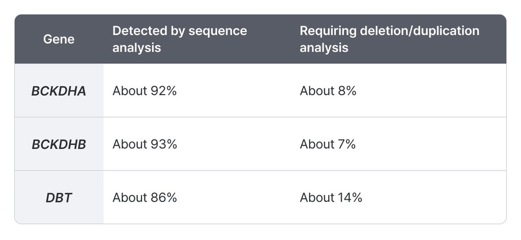
DBT in particular shows a relatively high proportion of pathogenic deletions.
This matters for the molecular diagnosis of MSUD. When analysis of single-nucleotide variants (SNVs) and small insertions/deletions (indels) alone does not adequately account for the genetic cause, copy number variants (CNVs) including single-exon or multi-exon deletions also have to be considered.
In an autosomal recessive disorder especially, when only one pathogenic variant is identified, it is necessary to consider a CNV on the other allele or a variant that is difficult to detect by standard sequence analysis.
6. Genotype and Clinical Severity
6-1. Does the causative gene determine MSUD severity?
Associations between causative gene and clinical severity have been reported in MSUD. These relationships are not absolute, however, and variants in the same gene can produce a range of clinical phenotypes.
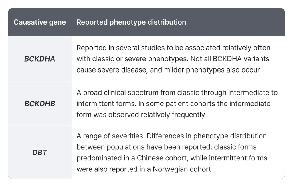
These findings show the limits of predicting disease severity from the causative gene alone.
Understanding clinical severity in MSUD requires considering not only which gene carries the variant, but also how that variant affects protein function and residual enzyme activity.
6-2. Genotype–Phenotype Correlation: Variant Type and Residual Enzyme Activity
In MSUD, genotype–phenotype correlation relates mainly to how much a variant reduces the function of the BCKD complex.
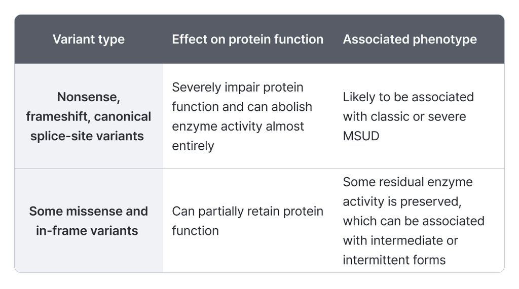
Variants associated with relatively mild or intermittent phenotypes are listed below.
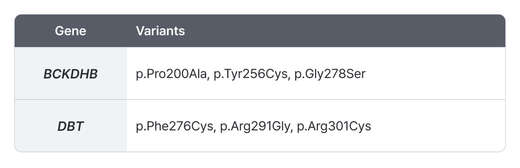
The fact that a given variant was reported in a patient with a mild phenotype is not by itself enough to conclude that every patient carrying that variant will have a mild clinical course. Even among patients with the same genotype, the frequency of metabolic crises and the long-term neurological outcome can differ.
A patient’s clinical course is influenced by many factors, including the timing of diagnosis, the state of metabolic control, the frequency and severity of metabolic crises, and the timing and effect of treatment.
In the genetic interpretation of MSUD, therefore, assessing the pathogenicity of a variant is one question, and judging how far that variant predicts clinical severity is a separate one that calls for care.
Conclusion
Maple syrup urine disease (MSUD) is a rare inherited metabolic disorder caused by reduced function of the BCKD complex, which acts in branched-chain amino acid metabolism.
The main causative genes are BCKDHA, BCKDHB and DBT, and biallelic pathogenic variants in these genes lead to the accumulation of BCAAs and BCKAs, which can result in a range of neurological abnormalities.
Leucine accumulation in particular plays an important role in neurotoxicity and the development of metabolic encephalopathy, and a rise in alloisoleucine is a useful biochemical marker for diagnosis.
MSUD spans a broad clinical spectrum, from the classic form with a fatal course in the neonatal period to the intermittent form with relatively normal development at baseline.
These phenotypic differences are determined not only by the causative gene but also by the functional effect of the variant, residual enzyme activity, and the patient’s state of metabolic control.
Accurate diagnosis and interpretation of MSUD therefore calls for an approach that evaluates clinical symptoms, biochemical testing and molecular genetic analysis together.
In interpretation work like this, where several causative genes are in play and a case can rest on a single identified allele, GEBRA organizes the candidate variants across the BCKD complex genes so they can be reviewed on one screen.
How are you handling rare disease variant interpretation?
See the GEBRA interpretation workspace for yourself, free.
References
- Menkes, J. H., Hurst, P. L., & Craig, J. M. (1954). A new syndrome: Progressive familial infantile cerebral dysfunction associated with an unusual urinary substance. Pediatrics, 14(5), 462–467.
- Chuang, D. T., Chuang, J. L., & Wynn, R. M. (2006). Lessons from genetic disorders of branched-chain amino acid metabolism. The Journal of Nutrition, 136(1 Suppl), 243S–249S.
- Morton, D. H., Strauss, K. A., Robinson, D. L., Puffenberger, E. G., & Kelley, R. I. (2002). Diagnosis and treatment of maple syrup disease: A study of 36 patients. Pediatrics, 109(6), 999–1008.
- Nellis, M. M., et al. (2003). Relationship of causative genetic mutations in maple syrup urine disease with their clinical expression. Molecular Genetics and Metabolism, 80(1–2), 189–195.
- Schadewaldt, P., Bodner-Leidecker, A., Hammen, H. W., & Wendel, U. (1999). Significance of L-alloisoleucine in plasma for diagnosis of maple syrup urine disease. Clinical Chemistry, 45(10), 1734–1740.
- Frazier, D. M., et al. (2014). Nutrition management guideline for maple syrup urine disease: An evidence- and consensus-based approach. Molecular Genetics and Metabolism, 112(3), 210–217.
- Strauss, K. A., Puffenberger, E. G., & Carson, V. J. (2020). Maple syrup urine disease. GeneReviews®. University of Washington, Seattle.
- Strauss, K. A., et al. (2010). Classical maple syrup urine disease and brain development: Principles of management and formula design. Molecular Genetics and Metabolism, 99(4), 333–345.
Full series
View all