



Episode 14: The Cause of Inherited Macular Degeneration and ABCA4

Haewon Park
Clinical Genomics Scientist
I explore how insights from genetic data can inform patient diagnosis and make complex concepts in genomic medicine accessible to a broader audience.
When the center of the visual field gradually blurs and reading or recognizing faces becomes difficult, an abnormality of the macula — the central region of the retina — may be suspected. A number of conditions affect the macula, and among them Stargardt disease is a representative inherited macular degeneration that begins in childhood or adolescence.
In this Clinical Series, we look at the main clinical features of Stargardt disease, along with how variants in the ABCA4 gene lead to lipofuscin accumulation in the retina and, ultimately, to vision loss.

FAQs
Q.How is Stargardt disease inherited?
A. Its most representative form, Stargardt disease 1 (STGD1), is inherited in an autosomal recessive manner, and generally occurs when disease-causing variants are present on both ABCA4 alleles.
Q. Why do the age of onset and rate of progression differ between patients?
A. Symptoms usually begin in childhood or adolescence, but the actual age of onset and the rate of progression vary considerably between patients. The type of ABCA4 variant and its residual function may also contribute to this clinical variability.
Q. Does peripheral vision also deteriorate in the early stages?
A. In the early stages of the disease, the peripheral retina is relatively preserved, and the peripheral visual field is often maintained within normal limits. What is affected first is the macula, at the center of the retina.
Q. What findings are observed on fundus examination and imaging?
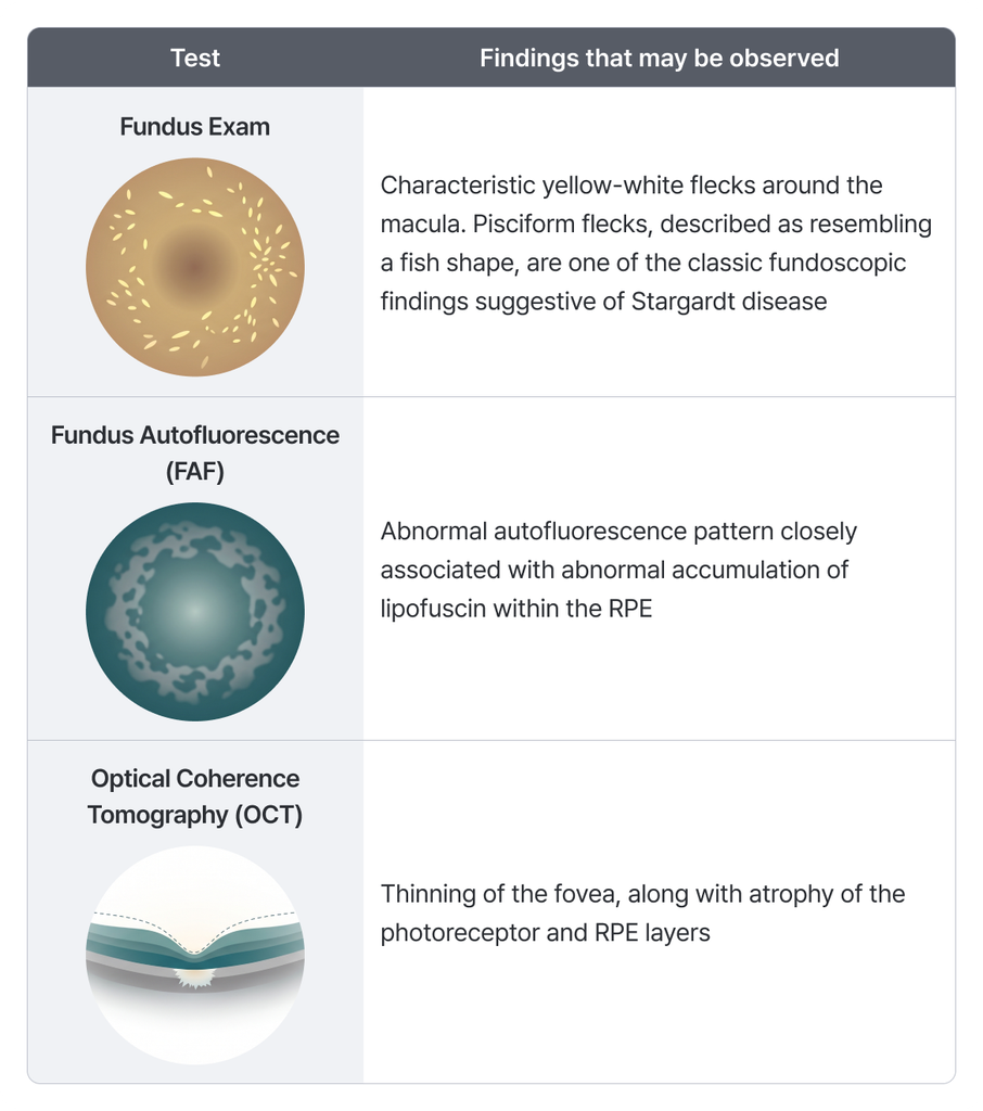
A. On fundus examination, characteristic yellow-white flecks may be seen around the macula; pisciform flecks, so named for their resemblance to the shape of a fish, are one of the representative fundus findings suggestive of Stargardt disease. On fundus autofluorescence (FAF), hyperautofluorescent flecks related to lipofuscin accumulation, together with hypoautofluorescence in areas of retinal pigment epithelium (RPE) atrophy, may be observed. On OCT, thinning of the fovea may be seen along with atrophy of the photoreceptor and RPE layers.
Q. How does genetic testing help in Stargardt disease?
A. Beyond distinguishing Stargardt disease from other clinically similar inherited retinal diseases, genetic testing can provide important information for confirming ABCA4 variants, understanding the mode of inheritance, and providing genetic counseling to patients and their families.
1. What is Stargardt disease?
Stargardt disease is an inherited retinal disease caused primarily by pathogenic variants in the ABCA4 gene. Its most representative form, Stargardt disease 1 (STGD1), is inherited in an autosomal recessive manner, and generally occurs when disease-causing variants are present on both ABCA4 alleles.
Symptoms usually begin in childhood or adolescence, but the actual age of onset and the rate of disease progression vary considerably between patients.
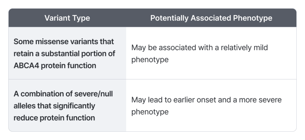
Variant type and residual function influence phenotype
The type of ABCA4 variant and its residual function may also contribute to this clinical variability.
2. Stargardt disease affects central vision first
The hallmark feature of Stargardt disease is progressive central vision loss.
As the macula at the center of the retina — particularly the photoreceptors and the retinal pigment epithelium (RPE) — is gradually damaged, the ability to distinguish fine detail, read text, and recognize faces may decline.
In the early stages of the disease, by contrast, the peripheral retina is relatively preserved, and the peripheral visual field is often maintained within normal limits.
- Findings on examination
So how does an abnormality in ABCA4 produce this characteristic pattern of retinal damage?
3. ABCA4: an important transporter in photoreceptors
ABCA4 is a large ATP-binding cassette transporter composed of approximately 2,273 amino acids. The ABCA4 protein is located in the disc membrane of the photoreceptor outer segment, where it plays an important role in maintaining the normal visual cycle through several functional domains.
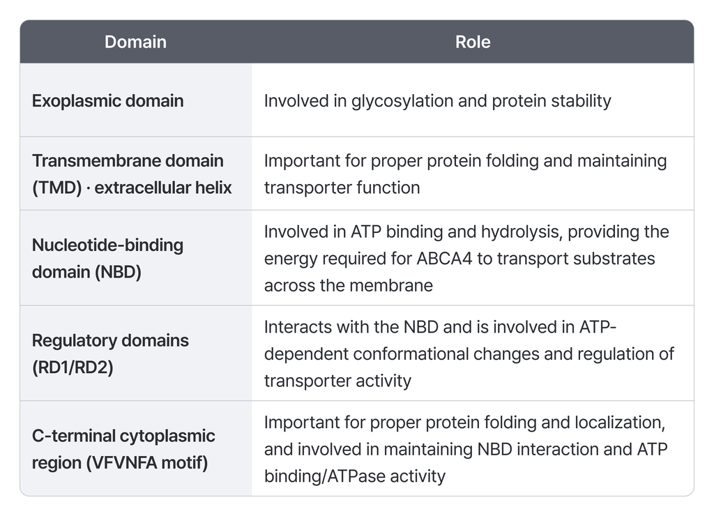
Variants arising in different regions of ABCA4 may therefore have varying effects on protein folding, expression, ATPase activity, or substrate transport, and consequently lead to differences in phenotype between patients.
4. The visual cycle that sustains vision
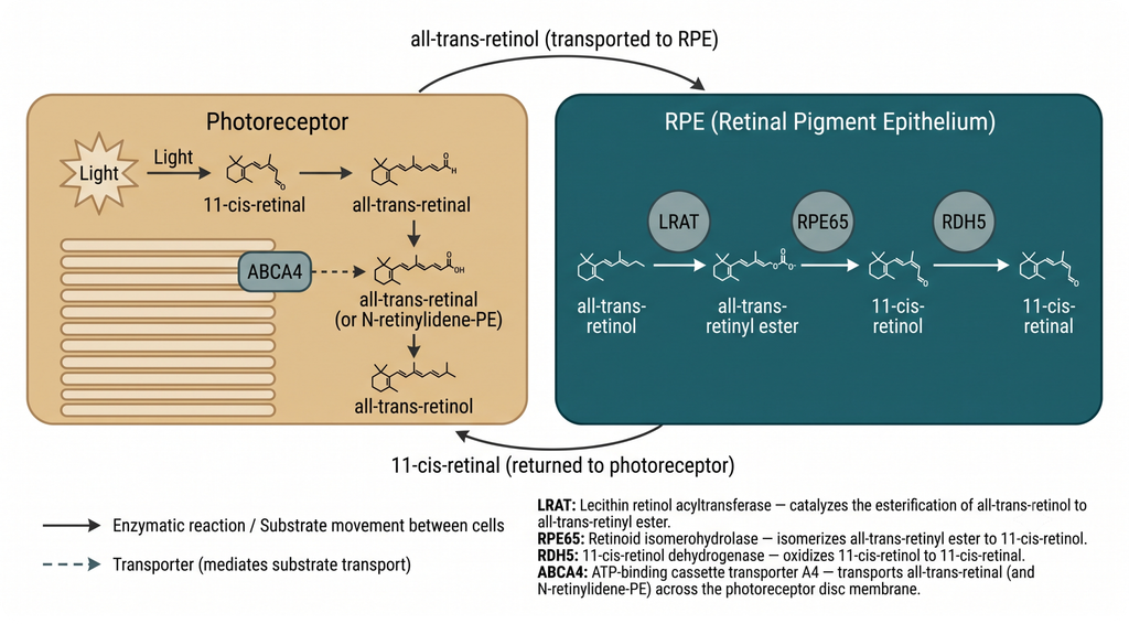
In photoreceptors that have detected light, 11-cis-retinal is converted to all-trans-retinal. This process is central to phototransduction, by which we perceive light, but the all-trans-retinal that is generated must then be processed.
Retinoids produced in photoreceptors are recycled through the visual cycle between the RPE and the photoreceptors. In the RPE, proteins such as LRAT, RPE65, and RDH5 participate in regenerating retinoids back into the 11-cis-retinal form, which is then used to form new photopigment in the photoreceptors.
Within this process, ABCA4 plays an important role in helping to clear the all-trans-retinal remaining in the photoreceptor disc.
5. What happens when ABCA4 does not work properly?
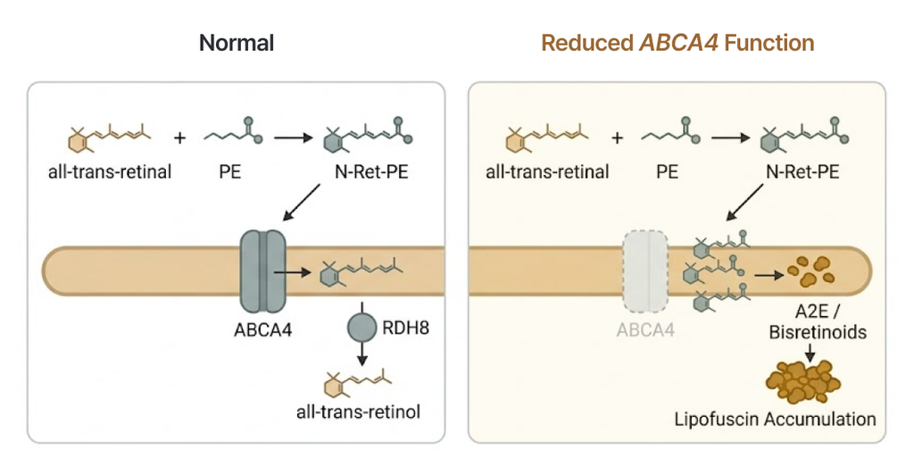
a. Under normal conditions
Inside the photoreceptor disc, all-trans-retinal can combine with phosphatidylethanolamine (PE) to form N-retinylidene-phosphatidylethanolamine (N-Ret-PE).
Normal ABCA4 uses ATP to help move N-Ret-PE from the lumenal leaflet to the cytoplasmic leaflet of the disc membrane. Once N-Ret-PE dissociates into all-trans-retinal and PE, the all-trans-retinal can be reduced to all-trans-retinol by enzymes such as RDH8, allowing it to re-enter the normal retinoid cycle.
b. When function is reduced
When ABCA4 function is reduced, N-Ret-PE and retinal-derived compounds are not sufficiently cleared. The accumulated retinal derivatives undergo a series of reactions to form bisretinoid compounds such as A2E, which accumulate in the RPE and become a major component of lipofuscin.
In the RPE of patients with Stargardt disease, markedly more lipofuscin may accumulate than normal.
6. From lipofuscin accumulation to vision loss
Excessive accumulation of lipofuscin — including bisretinoids such as A2E — does not simply end with waste building up inside the cell. The accumulation of these substances can interfere with normal RPE function and increase oxidative stress and cytotoxicity. Over time the RPE is damaged, and the photoreceptors, which depend on RPE function, may also be progressively lost.
The following sequence ultimately explains the core pathophysiology of Stargardt disease.
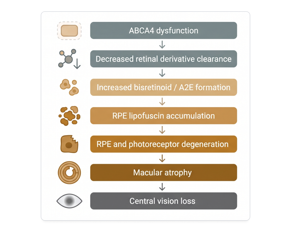
The characteristic yellow-white flecks observed on fundus examination are likewise related to this abnormal accumulation of lipofuscin and bisretinoids.
7. Stargardt disease, explained by a genetic variant
Stargardt disease is a genetic disease that clearly illustrates how an abnormality in a single gene can affect the metabolic processes of a specific tissue and ultimately lead to a characteristic clinical phenotype.
ABCA4 plays an important role in processing the retinal derivatives that arise during the visual cycle in photoreceptors. When this function is reduced, bisretinoids and lipofuscin accumulate abnormally in the RPE and, over the long term, cause degeneration of the photoreceptors and the RPE.
What genetic testing provides
Genetic testing in Stargardt disease can therefore provide the following information:
- Differentiation from other clinically similar inherited retinal diseases
- Confirmation of the ABCA4 variants causing Stargardt disease
- Determination of variant phase and inheritance pattern through family testing
- Genetic counseling for patients and their families
Closing
Behind the characteristic central vision loss and macular changes of Stargardt disease lies, ultimately, an intricate process in which a single small transporter in the photoreceptor maintains the normal visual cycle.
This is why ABCA4 and the visual cycle should be examined together when seeking to understand Stargardt disease.
How are you handling rare disease variant interpretation?
See the GEBRA interpretation workspace for yourself, free.
Reference
Abalem MF, Otte B, Andrews C, et al. Peripheral visual fields in ABCA4 Stargardt disease and correlation with disease extent on ultra-widefield fundus autofluorescence. Am J Ophthalmol. 2017.
Strauss RW, Kong X, Ho A, et al. Progression of Stargardt disease as determined by fundus autofluorescence over a 12-month period: ProgStar Report No. 11. JAMA Ophthalmol. 2019.
Heath Jeffery RC, Chen FK. Stargardt disease: Multimodal imaging: A review. Clin Exp Ophthalmol. 2021.
Curtis SB, Molday LL, Garces FA, Molday RS. Functional analysis and classification of homozygous and hypomorphic ABCA4 variants associated with Stargardt macular degeneration. Hum Mutat. 2020.
Al-Khuzaei S, Broadgate S, Foster CR, et al. An overview of the genetics of ABCA4 retinopathies, an evolving story. Genes (Basel). 2021.
Zhong M, Molday LL, Molday RS. Role of the C terminus of the photoreceptor ABCA4 transporter in protein folding, function, and retinal degenerative diseases. J Biol Chem. 2009.
Scortecci JF, Garces FA, Mahto JK, Molday LL, Van Petegem F, Molday RS. Structural and functional characterization of the nucleotide-binding domains of ABCA4 and their role in Stargardt disease. J Biol Chem. 2024.
Tripathy K, Kaur K. Stargardt Disease. StatPearls. 2024.
Molday RS, Garces FA, Scortecci JF, Molday LL. Structure and function of ABCA4 and its role in the visual cycle and Stargardt macular degeneration. Prog Retin Eye Res. 2022.
Giacalone JC, Scruggs BA. The pathology of Stargardt disease and future treatment horizons. Retinal Physician. 2024.
Full series
View all