[Interpretation Note #4] Interpreting CNVs and Deciding Whether to Report Them: From Transcript Impact to Dosage Sensitivity

Medical Genetics Group
3billion
We contribute to the discovery of causative variants in rare diseases through patient genome analysis, while strengthening clinical interpretation and research capabilities.
Copy number variants (CNVs) are deletions or duplications ranging from part of a single exon to several megabases. They are now routinely called from exome and genome data alongside SNVs, but they are interpreted differently. For a small intragenic CNV, the central question is how the structural change affects the transcript and gene function. For a large CNV, the central question is whether the change in gene dosage contributes to disease.
This article walks through the main steps a clinical interpreter may consider when working through a CNV, following the order of an actual analysis. From characterizing the CNV to deciding whether to report it, it sets out a reasonable analytical approach that can serve as a reference in clinical practice.

FAQs
Q1. Is every deletion of a known disease gene pathogenic?
A. No. Pathogenicity can vary depending on whether loss of function is the known disease mechanism for that gene, which part of the gene the deletion covers, how it affects the transcript, and the inheritance pattern of the condition.
Q2. Why check whether an intragenic deletion is in frame?
A. An out-of-frame deletion can shift the reading frame and create a premature stop codon, and if this triggers nonsense-mediated decay (NMD), the allele may lose function. An in-frame deletion preserves the reading frame, but its effect on protein function varies with the region removed.
Q3. What do the ClinGen HI and TS scores mean?
A. ClinGen’s dosage sensitivity scores indicate the level of evidence that a decrease (haploinsufficiency, HI) or increase (triplosensitivity, TS) in copy number of a given gene or genomic region can cause disease. HI and TS are assessed independently, and the scores are 0 (no evidence), 1 (limited evidence), 2 (emerging evidence) and 3 (sufficient evidence). Two separate special values exist: 30 indicates an association with an autosomal recessive condition, and 40 indicates evidence that dosage sensitivity is unlikely.
Q4. Is a CNV found in gnomAD or DGV automatically benign?
A. No. Some recurrent CNVs are found in healthy individuals but occur at higher frequency in patient cohorts. Presence in controls indicates incomplete penetrance, not that the CNV is harmless.
Q5. Can I use the breakpoints a read-depth caller reports as they are?
A. Read-depth based CNV callers often estimate the interval in which the copy number change occurred rather than the exact breakpoints. So when the position and extent of a CNV matter for interpreting its effect on the transcript or on gene function, account for breakpoint uncertainty and carry out additional confirmation as needed.
1. Define the CNV: deletion or duplication, and where?
Start by establishing the CNV type and the genes and genomic region affected. For a deletion, assess the possibility of haploinsufficiency (HI) from the reduced copy number; for a duplication, the possibility of triplosensitivity (TS) from the increased copy number. A duplication, however, may also disrupt gene function depending on breakpoint position and structure, not only through the increase in copy number. The ACMG/ClinGen guidelines therefore separate copy-number loss and gain and assess each against its own criteria.1)
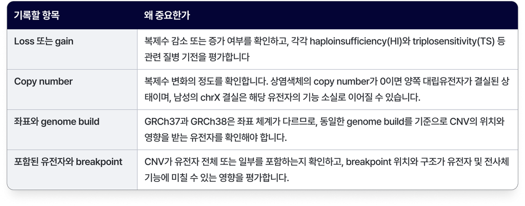
When a CNV lies within a gene, assessment centers on its effect on transcript structure and gene function. When it covers a whole gene or spans several genes, dosage sensitivity becomes the main consideration. In either case, transcript impact and dosage sensitivity may be reviewed together, depending on the structure of the CNV and the known disease mechanism.
2. Intragenic CNVs: predict the effect on the transcript
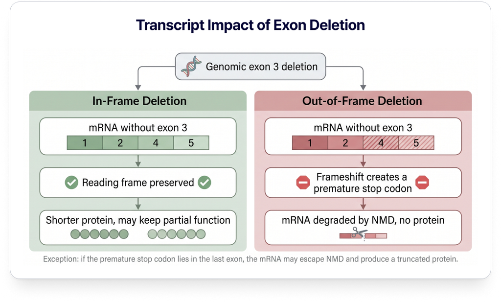
For a CNV with both breakpoints inside the same gene, what matters is assessing how the variant affects transcript structure and function. In practice, three questions guide this.
- Is the reading frame preserved? Check whether the length of coding sequence removed from or added to the actual transcript is a multiple of 3. Note that the effect can differ depending on splicing changes or the structure of the CNV.
- Is a premature termination codon (PTC) created? Assess whether a change in reading frame, or another mechanism, places a PTC upstream of the normal stop codon.
- Is nonsense-mediated decay (NMD) predicted? NMD is generally predicted when the PTC lies more than about 50–55 nucleotides upstream of the last exon–exon junction. Exceptions can occur depending on the position of the PTC and the structure of the transcript.
2-1. Examples
The examples below assume a hypothetical gene, Gene-X, and its transcript, and are for illustration. In real cases, the exon structure and breakpoints have to be confirmed.
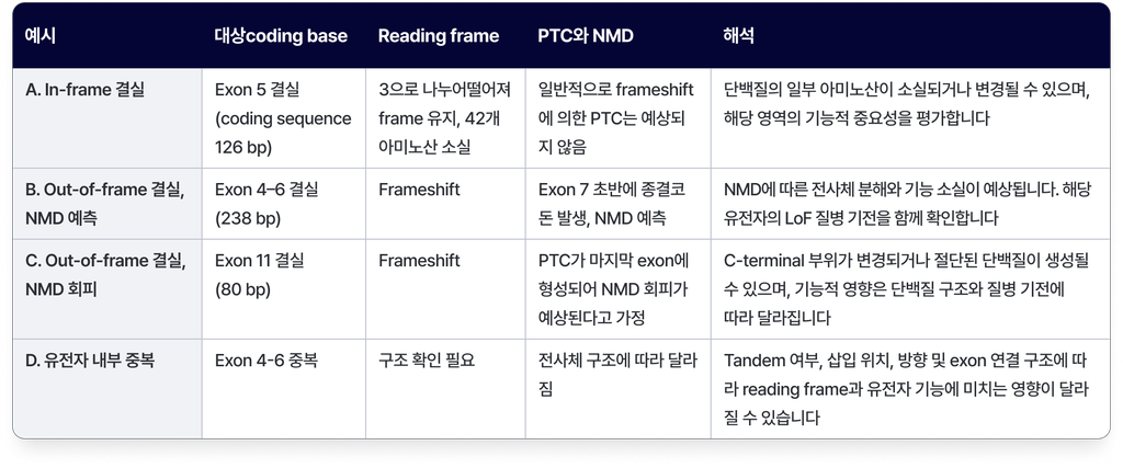
Read-depth based CNV callers often estimate the region in which the copy number change occurred rather than pinpointing exact breakpoints. So when assessing how the position and extent of a CNV affect transcript and gene function, account for breakpoint uncertainty and confirm by additional analysis where necessary.
3. Large or multigenic CNVs: check ClinGen dosage sensitivity
For a CNV covering a whole gene or a genomic region containing several genes, the assessment is whether a one-copy decrease or increase in that gene or region can cause disease. ClinGen Dosage Sensitivity curation presents this as two separate scores. 4) 5)
3-1. The HI and TS scores
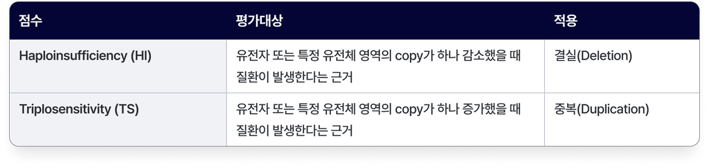
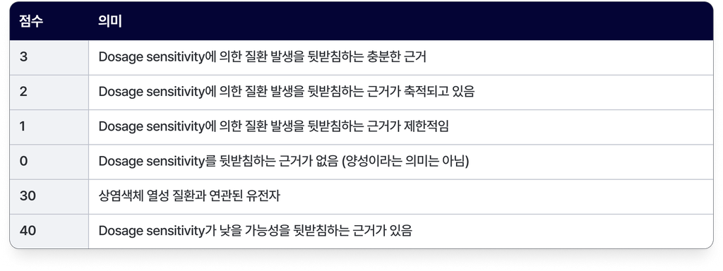
For example, USH1C is associated with autosomal recessive Usher syndrome and is scored by ClinGen as HI 30 and TS 0. A heterozygous whole-gene deletion of USH1C therefore generally suggests carrier status rather than disease. Disease can still occur, however, when a pathogenic variant is present on the other allele.
3-2. Assess at two levels
- Gene level: Check the HI score (for deletions) or TS score (for duplications) of the genes contained in the CNV. If an established dosage-sensitive gene is fully contained in the CNV, the copy number change in that gene may explain the patient’s phenotype. The inheritance pattern and the patient’s clinical features still have to be considered.
- Region level: Genomic regions associated with recurrent microdeletion and microduplication syndromes are curated at the region level. Check whether the CNV contains the entire region, or the known causative genes or critical region within it. Where the overlap is partial, the strength of the evidence varies with the extent of overlap and the genes included.
3-3. What to check on the curation page
The ClinGen Dosage Sensitivity website shows HI and TS scores by gene and by genomic region. Each curation page shows the supporting literature (PubMed IDs), the associated condition, genomic coordinates (GRCh37/GRCh38), constraint metrics such as pLI and LOEUF, and the date of the last evaluation.
The date of the last evaluation is particularly worth checking. A score of 0 assigned long ago may mean that evidence for dosage sensitivity was insufficient at the time of evaluation, and does not guarantee that no evidence exists today.
4. No ClinGen curation: search the literature and databases
Many CNVs fall in regions without ClinGen curation. In that case, build the evidence from published cases, which corresponds to Section 4 of the ACMG/ClinGen framework.1) Search by cytoband and CNV type. For a deletion at 1q24q25, for example, try “1q24q25 microdeletion syndrome” and “1q24q25 microdeletion”, then move on to narrower bands and the main genes in the region. PubMed, DECIPHER, ClinVar and OMIM are good starting points.
When comparing against published CNV cases, assess the following two things together.
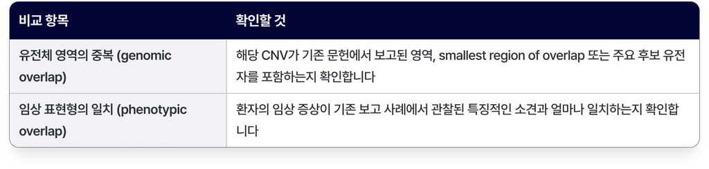
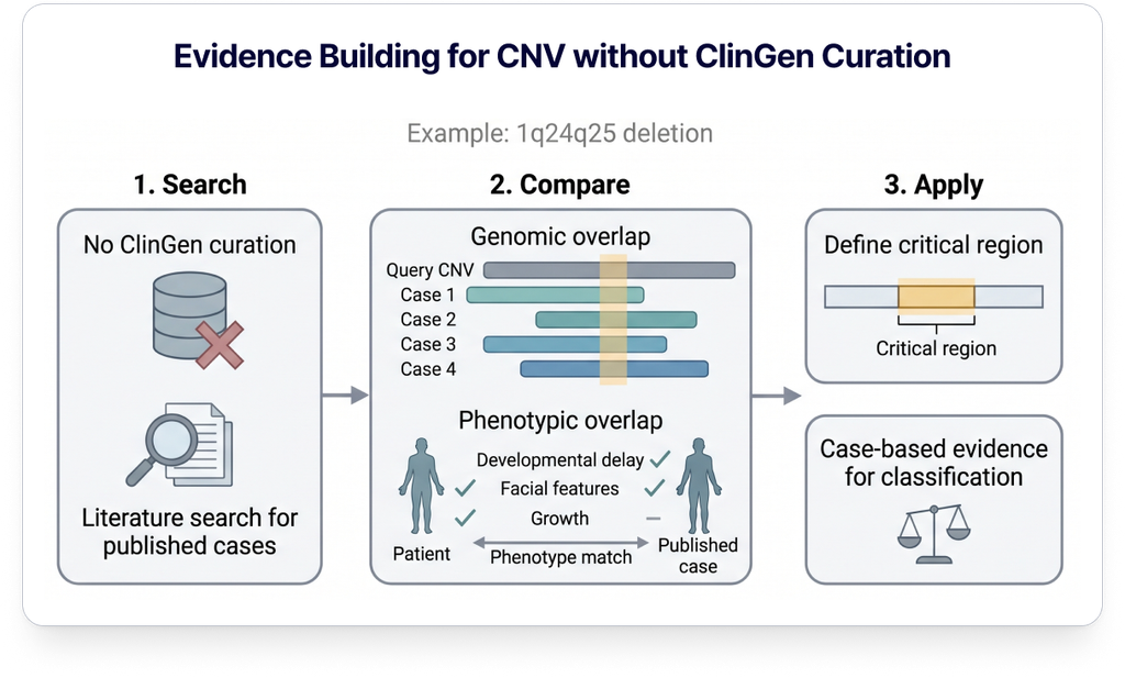
For example, Burkardt et al. (2011) reported growth retardation, microcephaly, small hands and feet with brachydactyly, and intellectual disability in nine patients with a 1q24q25 deletion, and proposed a critical region of approximately 1.9 Mb within 1q24.3q25.1. If a patient’s deletion overlaps that region and the clinical features are similar, the published literature can be used as evidence supporting the clinical relevance of the CNV. Where only part of the region overlaps, or the phenotype does not match closely enough, a more cautious interpretation is needed.
Bear in mind as well that older array-based studies may not have established the exact boundaries of a CNV, and that published case reports may not fully capture the range of phenotypes.
5. Caveat: recurrent CNVs with low penetrance
Some disease-associated CNVs show incomplete penetrance and can be found in people without symptoms. The 15q11.2 BP1–BP2 deletion is a well-known example: associations with developmental delay and learning and behavioural problems have been reported, but it is also observed in the general population and is known to have low penetrance.
5-1. Check population frequency
When assessing the pathogenicity of a CNV, check its frequency in the general population together with published cases. Databases such as DGV (Database of Genomic Variants) and gnomAD-SV can be used to check whether similar CNVs are observed in the general population.
Rather than simply looking at whether the genomic regions overlap, compare the following together.
- CNV type (deletion or duplication)
- Size
- Genes contained
- Extent of overlap
5-2. Before calling a CNV benign
The fact that a CNV is observed in the general population, or was inherited from an unaffected parent, is not by itself grounds for calling it benign. For CNVs with low penetrance in particular, also review how well the patient’s clinical phenotype fits and whether another causative variant may be present.
It is important to assess the pathogenicity classification of the CNV itself separately from whether it contributes to this particular patient’s phenotype.
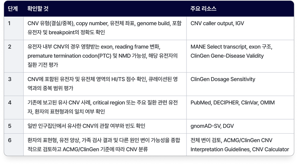
6. Practical checklist
Conclusion
CNV interpretation starts with establishing the type, position and size of the variant and the genes it contains. Intragenic CNVs are assessed by their effect on the transcript and protein function, while CNVs covering whole genes or several genes are assessed by the dosage sensitivity of those genes and genomic regions. Similar cases reported in the literature, population frequency, the patient’s clinical phenotype and the inheritance pattern all have to be weighed together in assessing the pathogenicity and clinical relevance of the CNV.
CNV interpretation is therefore more than establishing where a copy number change occurred. It is a process of reviewing gene structure and function, the evidence accumulated to date, and the patient’s clinical information together. GEBRA is a platform that supports this kind of CNV analysis and variant interpretation, and you can see it for yourself through a free trial.
How are you handling rare disease variant interpretation?
See the GEBRA interpretation workspace for yourself, free.
References
- Riggs ER, Andersen EF, Cherry AM, et al. Technical standards for the interpretation and reporting of constitutional copy-number variants: a joint consensus recommendation of the ACMG and ClinGen. Genet Med. 2020;22(2):245-257. PMID: 31690835.
- Abou Tayoun AN, Pesaran T, DiStefano MT, et al. Recommendations for interpreting the loss of function PVS1 ACMG/AMP variant criterion. Hum Mutat. 2018;39(11):1517-1524. doi: 10.1002/humu.23626.
- Newman S, Hermetz KE, Weckselblatt B, Rudd MK. Next-generation sequencing of duplication CNVs reveals that most are tandem and some create fusion genes at breakpoints. Am J Hum Genet. 2015;96(2):208-220. PMID: 25640679.
- ClinGen Dosage Sensitivity curation. search.clinicalgenome.org/kb/gene-dosage.
- Thaxton C, Good ME, DiStefano MT, et al. Utilizing ClinGen gene-disease validity and dosage sensitivity curations to inform variant classification. Hum Mutat. 2022. doi: 10.1002/humu.24291.
- ClinGen Dosage Sensitivity curation: USH1C. dosage.clinicalgenome.org.
- Burkardt DD, et al. Distinctive phenotype in 9 patients with deletion of chromosome 1q24-q25. Am J Med Genet A. 2011;155A(6):1336-1351. PMID: 21548129.
- Doornbos M, et al. Nine patients with a microdeletion 15q11.2 between breakpoints 1 and 2 of the Prader-Willi critical region, possibly associated with behavioural disturbances. Eur J Med Genet. 2009. PMID: 19328872.
- Rosenfeld JA, Coe BP, Eichler EE, Cuckle H, Shaffer LG. Estimates of penetrance for recurrent pathogenic copy-number variations. Genet Med. 2013;15(6):478-481. doi: 10.1038/gim.2012.164.
- Collins RL, Brand H, Karczewski KJ, et al. A structural variation reference for medical and population genetics. Nature. 2020;581(7809):444-451. PMID: 32461652.
- Minoche AE, Lundie B, Peters GB, et al. ClinSV: clinical grade structural and copy number variant detection from whole genome sequencing data. Genome Med. 2021;13:32. PMID: 33632298.
Full series
View all