



Episode 15: Homocystinuria — Causative Genes and How to Tell Them Apart

Medical Genetics Group
3billion
We contribute to the discovery of causative variants in rare diseases through patient genome analysis, while strengthening clinical interpretation and research capabilities.
In the body, methionine taken in through food is used across a number of metabolic processes. One of the important intermediate metabolites generated along the way is homocysteine.
Under normal circumstances, homocysteine is metabolized either through the remethylation pathway, in which it is converted back into methionine, or through the transsulfuration pathway, in which it passes through cystathionine and is converted into cysteine.
When the function of an enzyme or cofactor involved in these metabolic processes is impaired, however, homocysteine may not be processed normally and can accumulate in the body. The inherited metabolic disorders in which blood homocysteine concentrations rise markedly and homocysteine/homocystine is also increased in the urine are collectively known as homocystinuria.
FAQs
Q.If homocysteine is elevated, is it always CBS deficiency?
A. No. Homocystinuria can also occur when there is a problem in the remethylation pathway, which converts homocysteine back into methionine. In MTHFR deficiency, for example, reduced production of 5-methyltetrahydrofolate leads to impaired remethylation.
Q. How do you distinguish CBS deficiency from a remethylation disorder?
A. Methionine is the key clue. In CBS deficiency, transsulfuration is impaired, and it is characteristic for homocysteine and methionine to rise together. By contrast, in disorders related to MTHFR, MTR, MTRR and cobalamin metabolism, remethylation is impaired, so homocysteine rises while methionine may be low or within the normal range.
Q. Why should methylmalonic acid also be checked?
A. Because in disorders that affect both the adenosylcobalamin and methylcobalamin pathways — MMACHC among them — elevated methylmalonic acid may accompany the rise in homocysteine.
Q. Can a gene that does not directly encode a metabolic enzyme be a cause?
A. It can. HCFC1, for example, is associated with the regulation of MMACHC expression, and pathogenic variants can cause the X-linked cobalamin disorder Methylmalonic aciduria and homocystinuria, cblX type. In addition, variants in a specific region of PRDX1 can give rise to abnormal read-through transcription, which in turn can induce promoter methylation and transcriptional silencing of the adjacent MMACHC.
1. How is homocysteine metabolized?
Homocysteine metabolism proceeds in two broad directions.
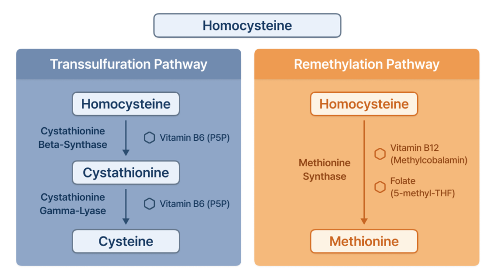
Severe hyperhomocysteinemia or homocystinuria can therefore arise not only from abnormalities in genes such as CBS, MTHFR, MTR and MTRR, but also from abnormalities in genes involved in the intracellular processing and trafficking of vitamin B12.
2. What problems arise when homocysteine accumulates?
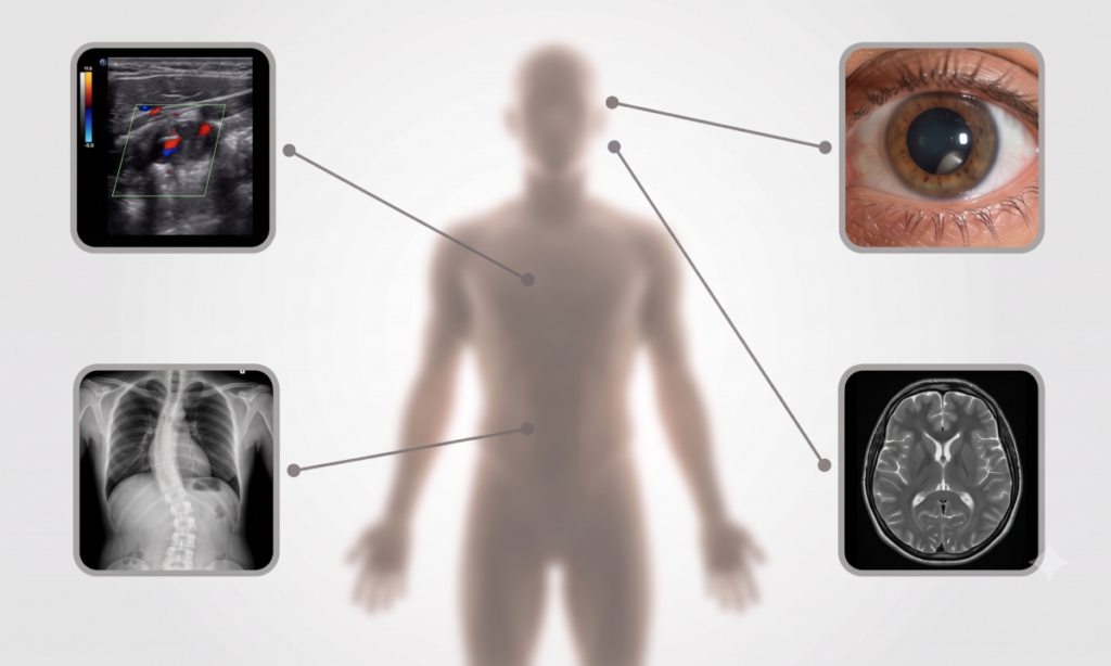
Excessive accumulation of homocysteine can affect a number of tissues.
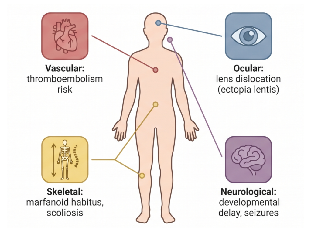
The combination and severity of symptoms differ considerably between patients. Homocystinuria is therefore better understood not as a disease confined to a single organ, but as a multisystem metabolic disorder that can produce a range of symptoms across the vascular, ocular, skeletal, and neurological systems.
3. The representative cause: CBS deficiency
The best-known form is cystathionine beta-synthase deficiency, caused by biallelic pathogenic variants in the CBS gene.
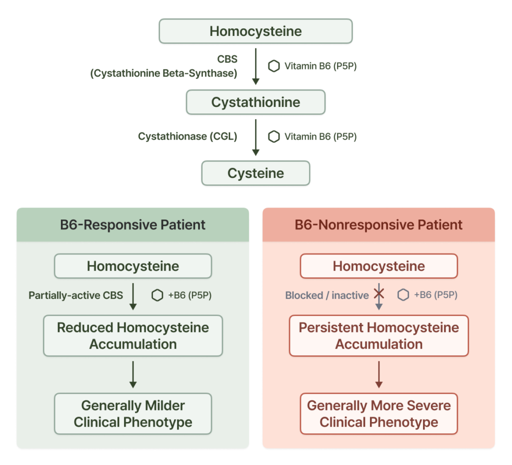
When CBS function is reduced, homocysteine is not normally converted into cystathionine. As a result, plasma total homocysteine rises markedly, and methionine generally rises as well.
CBS deficiency is an autosomal recessive disorder, and the clinical picture varies widely even within the same disease.
3-1. Pyridoxine responsiveness
One particularly important feature is pyridoxine (vitamin B6) responsiveness.
In some patients, administration of pyridoxine increases the residual activity of CBS, and homocysteine concentrations fall markedly. These B6-responsive patients generally tend to have a milder clinical picture than B6-nonresponsive patients, but actual responsiveness is best assessed through the response itself rather than concluded from genotype alone.
In patients with suspected CBS deficiency, it is therefore important to evaluate the pyridoxine response alongside biochemical findings and molecular genetic findings.
4. Not all homocystinuria is CBS deficiency
An elevated homocysteine does not always mean there is a problem with CBS. Homocystinuria can also occur when there is a problem in the remethylation pathway, which converts homocysteine back into methionine.
In MTHFR deficiency, for example, reduced production of 5-methyltetrahydrofolate leads to impaired remethylation. In this case, severe MTHFR deficiency causes a marked rise in homocysteine, while methionine generally falls.
“This is an important biochemical clue, in contrast to CBS deficiency, in which both homocysteine and methionine rise.”
Pathogenic variants in MTR and MTRR can likewise interfere with the methionine synthase system and cause homocystinuria. In these disorders too, impaired remethylation can produce hyperhomocysteinemia with low or normal methionine.
5. Causes related to cobalamin metabolism
Vitamin B12 plays an important role in homocysteine metabolism. Cobalamin that has entered the cell must be processed through several steps into an active cofactor.
When a problem arises in this process, methylcobalamin cannot be properly supplied to methionine synthase, and the remethylation of homocysteine may be impaired.
5-1. MMACHC and Methylmalonic aciduria and homocystinuria, cblC type
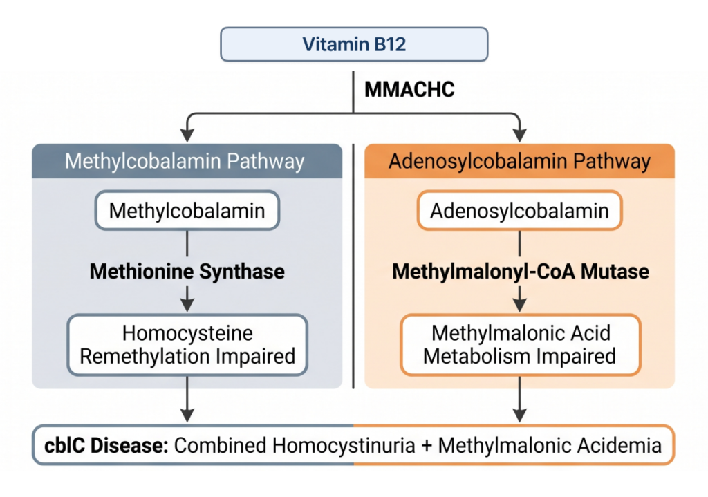
The representative gene is MMACHC. In MMACHC-related Methylmalonic aciduria and homocystinuria, cblC type, adenosylcobalamin metabolism as well as methylcobalamin can be affected, and it is therefore characteristic for homocystinuria and methylmalonic acidemia to appear together.
Patients may show developmental delay, neurologic abnormalities, hematologic abnormalities, ophthalmologic manifestations and abnormalities of various organs, and the clinical picture varies from infantile-onset to late-onset.
5-2. In the same MMADHC gene, phenotype differs by variant location
MMADHC is a particularly interesting gene in cobalamin metabolism. MMADHC pathogenic variants can produce different biochemical phenotypes depending on the location of the variant and its functional effect.
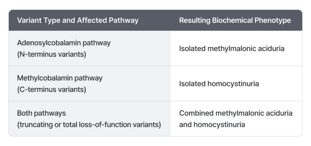
In MMADHC, then, what matters for understanding the phenotype is not simply “which gene carries the variant” but “where in the protein the variant sits and which function it impairs.”
5-3. Genes that are not direct metabolic enzymes can also be causative
The causes of homocystinuria and combined methylmalonic acidemia/homocystinuria are not necessarily limited to enzymes that directly catalyze homocysteine.
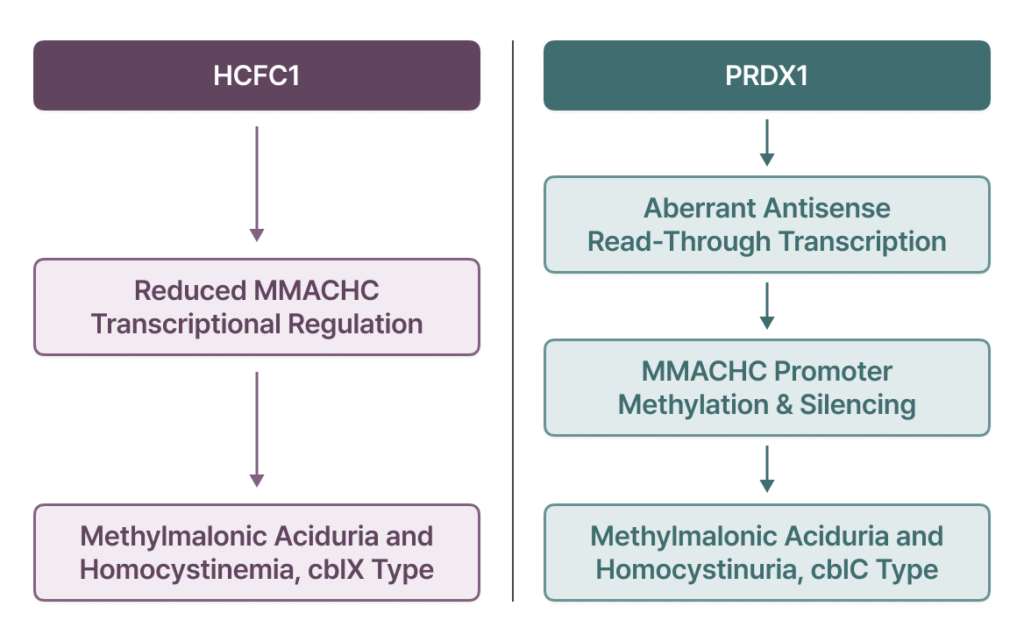
In metabolic disease, in other words, genetic abnormalities that alter gene expression and epigenetic regulation — not only the coding sequence of a particular enzyme — can also be the cause of disease.
6. How do you differentiate the causative pathway?
What matters in homocystinuria is that the underlying mechanism can differ even when the same biochemical phenotype of “homocysteine elevation” is seen.
- CBS deficiency — transsulfuration is impaired, and it is characteristic for homocysteine and methionine to rise together.
- MTHFR, MTR, MTRR and disorders related to methylcobalamin metabolism — remethylation is impaired, so homocysteine rises but methionine may be low or within the normal range.
- Disorders affecting both the adenosylcobalamin and methylcobalamin pathways, MMACHC among them — here methylmalonic acid elevation may accompany as well.
Rather than checking plasma total homocysteine alone, it is therefore very important to evaluate methionine and methylmalonic acid together in order to differentiate the cause.
6-1. Estimating the causative pathway from the biochemical profile
When severe hyperhomocysteinemia has been identified, the causative pathway can be estimated on the basis of the biochemical profile.
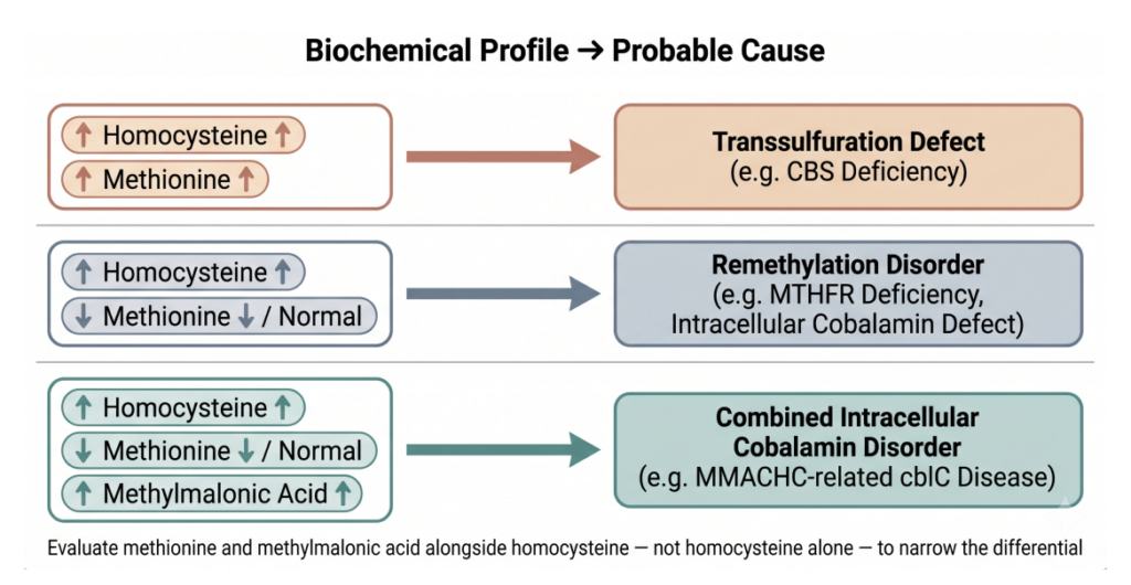
Interpreting these biochemical findings together with the patient’s clinical symptoms and genetic testing results allows a more accurate diagnosis to be approached.
Take-home message
Homocystinuria is not a single disease so much as an important biochemical phenotype that can appear across a number of genetic disorders affecting homocysteine metabolism.
The most representative cause is CBS deficiency, but genes in the remethylation pathway such as MTHFR, MTR and MTRR, as well as a range of genes related to intracellular cobalamin metabolism such as MMACHC, MMADHC and HCFC1, can also be causative.
Moreover, even with abnormalities in the same pathway or the same gene, onset, severity and the biochemical profile can differ according to the location of the variant and its residual function.
In patients with suspected homocystinuria, it is therefore important to evaluate not only the rise in homocysteine itself but also changes in methionine and methylmalonic acid, the clinical phenotype, and the underlying genetic cause, taken together.
This approach can provide an important clue for identifying the precise cause in complex homocysteine metabolism disorders and for deciding on an appropriate direction for diagnosis and treatment.
GEBRA is a platform that supports this kind of rare disease variant interpretation, and you can try it for yourself free of charge.
How are you handling rare disease variant interpretation?
See the GEBRA interpretation workspace for yourself, free.
References
Althubity, A. A. (2025). Homocystinuria: Advances in metabolic and molecular therapies targeting homocysteine pathways. Molecular Medicine Reports, 33(1), 34.
Esse, R., Barroso, M., Tavares de Almeida, I., & Castro, R. (2019). The contribution of homocysteine metabolism disruption to endothelial dysfunction: State-of-the-art. International Journal of Molecular Sciences, 20(4), 867.
Frosst, P., Blom, H. J., Milos, R., Goyette, P., Sheppard, C. A., Matthews, R. G., et al. (1995). A candidate genetic risk factor for vascular disease: A common mutation in methylenetetrahydrofolate reductase. Nature Genetics, 10(1), 111–113.
Goyette, P., Sumner, J. S., Milos, R., Duncan, A. M., Rosenblatt, D. S., Matthews, R. G., & Rozen, R. (1994). Human methylenetetrahydrofolate reductase: Isolation of cDNA, mapping and mutation identification. Nature Genetics, 7(2), 195–200.
Guéant, J. L., Chéry, C., Oussalah, A., Nadaf, J., Coelho, D., Josse, T., et al. (2018). A PRDX1 mutant allele causes a MMACHC secondary epimutation in cblC patients. Nature Communications, 9(1), 67.
Gupta, S., Kelow, S., Wang, L., Andrake, M. D., Dunbrack, R. L., & Kruger, W. D. (2018). Mouse modeling and structural analysis of the p.G307S mutation in human cystathionine β-synthase (CBS) reveal effects on CBS activity but not stability. Journal of Biological Chemistry, 293(36), 13921–13931.
Jakubowski, H., Zhang, L., Bardeguez, A., & Aviv, A. (2000). Homocysteine thiolactone and protein homocysteinylation in human endothelial cells: Implications for atherosclerosis. Circulation Research, 87(1), 45–51.
Koklesova, L., Mazurakova, A., Samec, M., Biringer, K., Samuel, S. M., Büsselberg, D., et al. (2021). Homocysteine metabolism as the target for predictive medical approach, disease prevention, prognosis, and treatments tailored to the person. EPMA Journal, 12(4), 477–505.
Kraus, J. P., Janošík, M., Kožich, V., Mandell, R., Shih, V., Sperandeo, M. P., et al. (1999). Cystathionine β-synthase mutations in homocystinuria. Human Mutation, 13(5), 362–375.
Lerner-Ellis, J. P., Anastasio, N., Liu, J., Coelho, D., Suormala, T., Stucki, M., et al. (2009). Spectrum of mutations in MMACHC, allelic expression, and evidence for genotype–phenotype correlations. Human Mutation, 30(7), 1072–1081.
Lerner-Ellis, J. P., Tirone, J. C., Pawelek, P. D., Doré, C., Atkinson, J. L., Watkins, D., et al. (2006). Identification of the gene responsible for methylmalonic aciduria and homocystinuria, cblC type. Nature Genetics, 38(1), 93–100.
Poddar, R., & Paul, S. (2009). Homocysteine–NMDA receptor-mediated activation of extracellular signal-regulated kinase leads to neuronal cell death. Journal of Neurochemistry, 110(3), 1095–1106.
Pizzorno, J. (2014). Homocysteine: Friend or foe? Integrative Medicine: A Clinician’s Journal, 13(4), 8.
Ruiz-Mercado, M., Vargas, M. T., de Soto, I. P., Pecellín, C. D., Sánchez, M. C., Delgado, M. B., et al. (2016). Methionine synthase reductase deficiency (CblE): A report of two patients and a novel mutation. Hematology, 21(3), 193–197.
Sacharow, S. J., & Levy, H. L. (2025). Homocystinuria due to cystathionine beta-synthase deficiency. In GeneReviews®.
Sauls, D. L., Wolberg, A. S., & Hoffman, M. (2003). Elevated plasma homocysteine leads to alterations in fibrin clot structure and stability: Implications for the mechanism of thrombosis in hyperhomocysteinemia. Journal of Thrombosis and Haemostasis, 1(2), 300–306.
Stucki, M., Coelho, D., Suormala, T., Burda, P., Fowler, B., & Baumgartner, M. R. (2012). Molecular mechanisms leading to three different phenotypes in the cblD defect of intracellular cobalamin metabolism. Human Molecular Genetics, 21(6), 1410–1418.
Torices, L., de Las Heras, J., Arango-Lasprilla, J. C., Cortés, J. M., Nunes-Xavier, C. E., & Pulido, R. (2021). MMADHC premature termination codons in the pathogenesis of cobalamin D disorder: Potential of translational readthrough reconstitution. Molecular Genetics and Metabolism Reports, 26, 100710.
Watkins, D., Ru, M., Hwang, H. Y., Kim, C. D., Murray, A., Philip, N. S., et al. (2002). Hyperhomocysteinemia due to methionine synthase deficiency, cblG: Structure of the MTR gene, genotype diversity, and recognition of a common mutation, P1173L. The American Journal of Human Genetics, 71(1), 143–153.
Yu, H. C., Sloan, J. L., Scharer, G., Brebner, A., Quintana, A. M., Achilly, N. P., et al. (2013). An X-linked cobalamin disorder caused by mutations in transcriptional coregulator HCFC1. The American Journal of Human Genetics, 93(3), 506–514.
Full series
View all