


VUS Reanalysis: What Actually Changes, and Who Can Change It

Sookjin Lee
Chief Business Officer (CBO) | Ph.D.
A business expert leading innovation in precision medicine and genomics for 20 years. Accelerating the commercialization of innovative technologies with academic expertise and market insight.
In brief
- A VUS is not a final answer. It means the evidence needed for a call is either insufficient or conflicting.
- Of the 28 ACMG criteria, only a few — PVS1, BA1, PS1 — rarely move. The other 20-plus can shift as evidence accumulates.
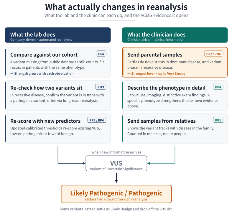
- The laboratory adds evidence through cohort recurrence, phase confirmation, and recalibrated predictors. The clinician adds evidence through parental samples, detailed phenotyping, and samples from relatives.
- The strongest single lever is a parental sample. In dominant disease it settles de novo status; in recessive disease it settles phase.
- Reanalysis is triggered by new information, not by elapsed time.
A first test sometimes ends with only a VUS (variant of uncertain significance), and the question that follows is practical: what can be done now?
Reanalysis is not a second look at the same data. It recalculates which ACMG criteria apply and at what strength. So the useful question is what new information changes that calculation, and who can generate it.
A VUS is an evidence gap, not a near-miss
The 2015 ACMG/AMP framework defines 28 criteria, each carrying a direction — pathogenic or benign — and a strength from supporting through very strong. A VUS means the applied criteria do not meet the combining rules, or that evidence points both ways. It does not mean “probably pathogenic.”
A few criteria are relatively stable: PVS1, BA1, and PS1 rest on protein-level or population-level facts that rarely reverse. The other 20-plus respond to new information, and that information comes from two different places.
What the laboratory can change
Recurrence in an accumulated cohort (PS4)
A variant with no entry in public databases still carries weight when it recurs in other patients with the same phenotype. In ultra-rare disease, where formal case-control comparison is rarely feasible, counting independent affected probands is the practical route to this criterion.
Phase of two variants (PM3)
In recessive disease, PM3 applies when the variant on the other allele is pathogenic or likely pathogenic; when that variant is itself a VUS, the strength drops considerably. ClinGen SVI sets strength from whether phase is confirmed and how the other variant is classified. Parental samples are the most direct route to phase; long-read sequencing can resolve it where parents are unavailable.
Recalibrated predictors (PP3 / BP4)
ClinGen SVI now recommends a single pre-specified tool applied at calibrated thresholds rather than a consensus of several. What separates laboratories is less which tool they run than whether thresholds are applied consistently and documented. When thresholds are updated, existing VUS are re-scored — in both directions.
What the clinician can change
Parental samples: one sample, two answers (PS2 / PM6)
A single request opens two distinct lines of evidence — in dominant disease, whether the variant arose de novo; in recessive disease, the phase of two variants, which raises PM3. De novo evidence has no fixed strength: ClinGen SVI scores it from confirmed parental relationships, phenotype consistency, and the number of de novo occurrences, reaching the very strong level where those conditions are met. De novo status alone does not settle pathogenicity; it is weighed with phenotype fit and population rarity.
Detailed phenotyping multiplies rather than adds (PP4)
Detailed reporting is often understood as adding PP4 as a separate line of evidence. UK ACGS guidance says otherwise: when PS2/PM6 applies, phenotype specificity should raise the strength of PS2/PM6 rather than be counted again under PP4. The implication is direct — parental samples and detailed phenotyping are multiplicative, not additive. Which is why “developmental delay” is not enough. Lab values, imaging findings, and distinctive examination findings, described at the granularity of HPO terms, are what create room for the call to change.
Samples from relatives (PP1)
Whether the variant tracks with disease through affected relatives is evidence for pathogenicity, counted in informative meioses rather than in people tested — and meioses can be summed across unrelated families. Samples from unaffected relatives serve a different function, entering the cosegregation calculation and the assessment of non-segregation (BS4).
When is a separate reanalysis request worth making?
The trigger is information, not time.
(O) Worth requesting — new clinical findings have emerged (new results, additional features, a refined diagnosis); parental or other family samples are now available; another investigation has produced a finding relevant to the diagnosis.
(X) Not usually necessary — the report is recent and none of the above has changed. Updates to public databases and new literature take time to reach the point of changing a specific call. Within roughly three months, a change driven only by those sources is uncommon, and where reanalysis runs automatically it should reach you when the change occurs.
Reanalysis does not only mean upgrades
Recalibration and updated frequency data work in the other direction too, and some variants settle as likely benign and drop off the follow-up list. That is also progress: a shorter candidate list means closer attention to what remains, and fewer families sent for surveillance that turns out not to be warranted.
A VUS is not a failed test. It marks evidence that has not yet accumulated, and much of that evidence is within reach of the laboratory and the clinician. Reanalysis is triggered by new information, not by elapsed time.
If a VUS in one of your patients could be reassessed — a parental sample that could now be added, or clinical findings that have emerged since the report — our clinical team is glad to look at the case with you.
Frequently asked questions
Q1. Can treatment proceed with a VUS result?
Treatment of the patient’s symptoms proceeds independently of variant classification. What a VUS constrains is any decision resting on the variant itself — recommending prophylactic measures to an asymptomatic relative on that basis, or determining carrier status from the variant alone.
Q2. Why does trio testing matter so much for diagnostic yield?
A variant that would remain a VUS on proband-only testing can gain strong evidence once parental testing shows it absent in both parents. In recessive disease, parental samples also establish whether two variants sit on different alleles.
Q3. Does detailed symptom reporting really change the classification?
It can. The strength of de novo evidence depends on how specific the phenotype is for the disorder in question, so lab values, imaging findings, and distinctive examination findings allow the same variant to carry more weight.
Q4. Why does the size of a laboratory’s in-house database affect classification?
Because a variant with no entry in public databases can still be shown to recur in other patients with the same phenotype. A patient database is not a control cohort, though — population frequency has to be assessed against separate ancestry-matched control data.
Q5. Do reanalysis results always move toward pathogenic?
No. Predictor recalibration and updated frequency data also work toward benign, and some variants are reclassified as likely benign and removed from follow-up.
References
- Richards S, Aziz N, Bale S, et al. Standards and guidelines for the interpretation of sequence variants: a joint consensus recommendation of the American College of Medical Genetics and Genomics and the Association for Molecular Pathology. Genet Med. 2015;17(5):405-424. doi:10.1038/gim.2015.30
- ClinGen Sequence Variant Interpretation Working Group. Recommendation for de novo Criteria (PS2/PM6), Version 1.1. PDF
- ClinGen Sequence Variant Interpretation Working Group. Recommendation for in trans Criterion (PM3), Version 1.0. PDF
- Pejaver V, Byrne AB, Feng BJ, et al. Calibration of computational tools for missense variant pathogenicity classification and ClinGen recommendations for PP3/BP4 criteria. Am J Hum Genet. 2022;109(12):2163-2177. doi:10.1016/j.ajhg.2022.10.013
- Association for Clinical Genomic Science. Best Practice Guidelines for Variant Classification in Rare Disease. PDF
- Jarvik GP, Browning BL. Consideration of cosegregation in the pathogenicity classification of genomic variants. Am J Hum Genet. 2016;98(6):1077-1081.
- ClinGen Sequence Variant Interpretation Working Group. Guidance for use of the PP1/BS4 co-segregation and PP4 phenotype specificity criteria. PMC10806742