



Red Blood Cells Shaped Like a Sickle? Understanding Sickle Cell Disease
Sickle cell disease is a group of inherited hemoglobinopathies caused by pathogenic variants in the HBB gene, which encodes the beta-globin subunit of adult hemoglobin.
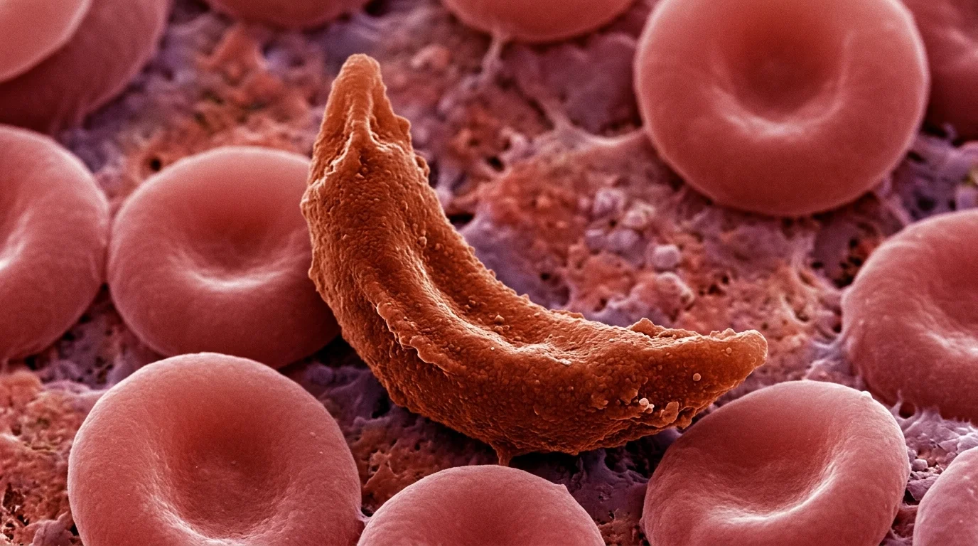
When two disease-causing HBB alleles are present, deoxygenated HbS polymerizes, distorting erythrocytes into rigid sickle shapes that drive hemolysis and vaso-occlusion.
Frequently asked questions
What is the difference between sickle cell trait and disease?
Sickle cell trait means carrying one pathogenic HBB allele; carriers are usually healthy. Sickle cell disease requires two disease-causing alleles and produces chronic hemolysis, vaso-occlusive episodes, and organ complications.
Why order genetic testing when electrophoresis is available?
Protein-based assays identify hemoglobin variants but can be ambiguous in newborns, after transfusion, or in compound heterozygotes. HBB sequencing resolves the precise genotype, supports carrier detection, and informs family and reproductive counseling.
What turns red blood cells into a sickle shape
The sickle variant is a single nucleotide change in HBB that swaps glutamic acid for valine in beta-globin. This substitution makes deoxygenated hemoglobin (HbS) polymerize, and the resulting fibers deform red cells into stiff sickle shapes.
Two processes then drive the clinical picture: chronic hemolytic anemia from shortened red cell lifespan, and vaso-occlusion as deformed cells block blood vessels. Vaso-occlusion leads to acute pain crises, acute chest syndrome, splenic sequestration, and organ damage that accumulates over time.
Prevalence and distribution
Sickle cell disease is among the most common monogenic disorders worldwide, with the highest allele frequencies in populations with historical malaria exposure. In the United States, the disease predominantly affects people of Sub-Saharan African descent, with an estimated prevalence of about 1 in 5000.
Carrier frequency is substantial. Roughly 2 million people carry sickle cell trait and about 72,000 live with sickle cell disease in the United States, underscoring the value of carrier screening in reproductive counseling.
How it’s detected and confirmed
Newborn screening detects most affected infants before symptoms appear, enabling early penicillin prophylaxis and parental education. A positive screen must be confirmed with follow-up testing.
Hemoglobin electrophoresis and HPLC quantify hemoglobin fractions and distinguish HbSS from HbSC or HbS/beta-thalassemia. These methods can be limited in the newborn period, after recent transfusion, or when a rare variant co-occurs.
Targeted HBB sequencing resolves these ambiguities. Molecular confirmation defines the exact genotype, identifies co-inherited thalassemia alleles, and provides the definitive result needed for cascade testing across a family.
* This article is educational and does not replace individualized medical advice. Diagnostic and management decisions should be made with a qualified clinician.
References
Sanyaolu A, et al. Current modalities of sickle cell disease management. 2020. https://doi.org/10.1097/BS9.0000000000000056

Soo-jung Baek
Marketing Manager
I strive to empower the rare disease community by sharing meaningful insights backed by our company’s expertise.