


Lissencephaly and LIS1/DCX Variants: What Genetic Testing Can Identify
Lissencephaly is a rare group of neuronal migration disorders characterized by the absence of normal convolutions (gyri) in the cerebral cortex, leading to a smooth or nearly smooth brain surface. This structural defect arises from a failure of neurons to migrate to their proper locations during the 12th to 24th weeks of gestation, resulting in a significantly thickened cortex with a simplified, four-layered structure instead of the normal six.
The Genetic Architecture of Lissencephaly
Normal brain development involves a highly orchestrated process where newly formed neurons migrate from their origin near the ventricles to the cerebral cortex. In lissencephaly, this process is disrupted. The two most significant genes implicated in classical lissencephaly are PAFAH1B1 (commonly known as LIS1) and DCX (Doublecortin).
LIS1-Associated Lissencephaly
The LIS1 gene, located on chromosome 17p13.3, was one of the first genes identified in relation to this condition. Mutations in LIS1 typically cause a pattern of lissencephaly that is more severe in the posterior regions of the brain. These mutations can range from point mutations to larger deletions, which can also encompass adjacent genes, leading to Miller-Dieker syndrome, a more severe condition with characteristic facial features and other birth defects.
DCX-Associated Lissencephaly
The DCX gene is located on the X chromosome and is associated with an X-linked form of lissencephaly. Due to its inheritance pattern, the phenotype differs significantly between sexes. In males, a pathogenic variant in DCX typically leads to classical lissencephaly, often with an anterior-predominant pattern. In heterozygous females, the same variant usually results in a milder phenotype known as subcortical band heterotopia (SBH), where a band of gray matter is misplaced beneath the cortex. For a female carrier, each pregnancy carries a 25% chance of having an affected son (a 50% chance of having a male child multiplied by a 50% chance he will inherit the pathogenic variant).
Other Genetic Causes
Together, mutations and deletions in LIS1 and DCX account for over 60% of isolated lissencephaly cases. However, a growing list of other genes has been identified in the remaining cases. These include ARX, RELN, and several tubulin genes like TUBA1A. These less common causes are associated with a range of lissencephaly subtypes, sometimes with additional features like agenesis of the corpus callosum or cerebellar hypoplasia.
Clinical Presentation and Diagnosis
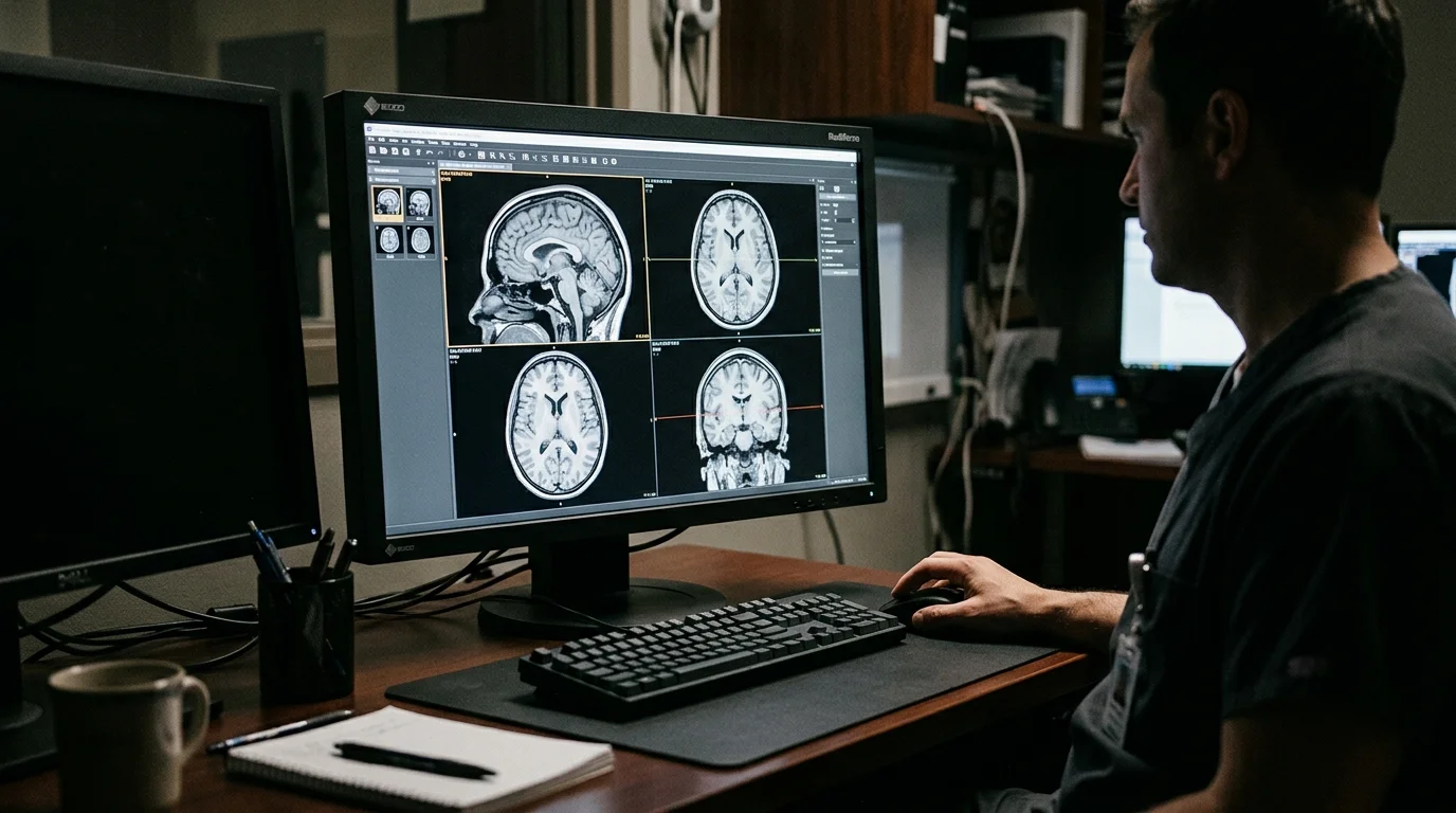
The diagnostic journey for lissencephaly begins with clinical suspicion in an infant presenting with developmental delay, abnormal muscle tone, and seizures. Neuroimaging is the next critical step.
Phenotypes and Classification
Lissencephaly is broadly grouped into two main categories based on histopathology. Classic lissencephaly (Type 1) is associated with a thickened, four-layered cortex and is typically caused by mutations in genes like LIS1 and DCX. In contrast, cobblestone lissencephaly (Type 2) involves a disorganized and nodular cortical surface and is associated with a different set of genes often linked to muscular dystrophies. As described in a review on the topic, Type 1 lissencephaly presents with a four-layered cortex instead of the normal six, while Type 2 appears disorganized and “pebbled.”
The Role of Genetic Testing
While an MRI can confirm the structural abnormality, genetic testing is essential to establish a definitive molecular diagnosis. This is important for several reasons:
- Prognostic Information: The specific gene and mutation can sometimes predict the severity of seizures and developmental outcome.
- Management: Identifying the genetic cause can help anticipate associated medical issues.
- Genetic Counseling: It provides families with accurate information about recurrence risk and allows for carrier testing of at-risk relatives.
Testing may begin with a targeted gene panel for known lissencephaly genes. For cases where panel testing is non-diagnostic, whole exome sequencing (WES) or whole genome sequencing (WGS) can be employed. This broader approach is effective in identifying variants in newly discovered genes or atypical presentations.
* This article is educational and does not replace individualized medical advice. Diagnostic and management decisions should be made with a qualified clinician.
Want to learn more about
3billion's genetic testing?
We'll reply within 1 business day.
References
- Pilz DT, et al. LIS1 and XLIS (DCX) mutations cause most classical lissencephaly, but different patterns of malformation. 1998. https://doi.org/10.1093/hmg/7.13.2029
- Haverfield EV, et al. Intragenic deletions and duplications of the LIS1 and DCX genes: a major disease-causing mechanism in lissencephaly and subcortical band heterotopia. 2009. https://doi.org/10.1038/ejhg.2008.213
- Jang MA, et al. Identification of DCX gene mutation in lissencephaly spectrum with subcortical band heterotopia using whole exome sequencing. 2013. https://doi.org/10.1016/j.pediatrneurol.2012.12.033
- Verloes A, et al. Genetic and clinical aspects of lissencephaly. 2007. https://doi.org/10.1016/s0035-3787(07)90460-9

Soo-jung Baek
Marketing Manager
I strive to empower the rare disease community by sharing meaningful insights backed by our company’s expertise.