


Mitochondrial Disease Doesn’t Arrive Looking Typical

Sookjin Lee
Chief Business Officer (CBO) | Ph.D.
A business expert leading innovation in precision medicine and genomics for 20 years. Accelerating the commercialization of innovative technologies with academic expertise and market insight.
Key findings
- We analyzed the pre-test clinical picture of 348 patients (38 countries, 2021–present) confirmed to carry pathogenic mtDNA variants.
- 286 (82%) had no documented elevated lactate, ragged-red fibers, or ophthalmoplegia.
- Among adults referred with suspected Fabry disease, 64 were diagnosed with mitochondrial disease — 56 with MELAS (MT-TL1).
- Three specialties accounted for 79% of cases: pediatric neurology (29%), ophthalmology (28%), cardiology (23%).
- For 75 LHON patients, optic atrophy was the only reported finding.
- 296 (85%) were diagnosed by exome-based testing. No separate mtDNA panel was performed.
The picture we wait for
Ask a clinician to describe mitochondrial disease and you’ll usually hear the same picture:
Elevated lactate. Ragged-red fibers on biopsy. A child with multi-organ involvement.
The problem is that mtDNA variants are rarely suspected until that picture appears. And when the picture never appears, there is no opportunity to find out whether the decision was right.
So we looked from the opposite direction. What did confirmed patients look like before testing?
1. 82% had no classic mitochondrial findings
Of 348 confirmed patients, 286 (82%) had no documented elevated lactate, ragged-red fibers, or ophthalmoplegia.
Four out of five were diagnosed without a single finding that would have raised suspicion.
Classic features are not a prerequisite. Used as a gate for testing, they exclude most of the patients who turn out to have the disease.
2. Fabry disease was suspected. It was MELAS.
The clearest example came from cardiology.
In a Korean multicenter program, exome testing was ordered for adults with suspected lysosomal storage disease — Fabry disease in particular. Presenting features were left ventricular hypertrophy, heart failure, proteinuria, tinnitus, and limb pain. A classic Fabry profile.
64 of them were diagnosed with mitochondrial disease instead. 56 carried MT-TL1 variants: MELAS.
None had lactate testing or muscle biopsy. Mitochondrial disease was never the question. Had these patients been worked up with GLA single-gene testing or enzyme assay, the result would have been negative, cause unknown.
Why the two overlap
m.3243A>G, which accounts for roughly 80% of MELAS, shares much of its clinical picture with Fabry disease.
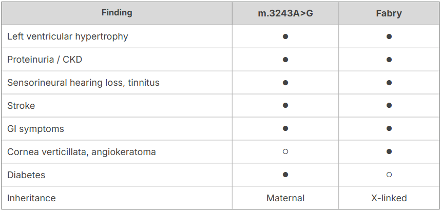
Five findings overlap. In clinic, LVH with proteinuria and tinnitus does not distinguish between them.
Two features help most:
- Pedigree. Maternal-only transmission points to mitochondrial disease. Father-to-son transmission rules it out.
- Diabetes with hearing loss. Absent in Fabry, and central to MIDD — another presentation of the same m.3243A>G variant.
One variant, several diagnoses
The same variant presents as cardiomyopathy, MELAS, or diabetes depending on which tissue carries the higher mutant load. Each cell holds hundreds to thousands of mitochondria, and the proportion carrying the variant — heteroplasmy — differs by tissue.
Mitochondrial disease is not confined to one specialty. The same variant is being diagnosed under different names in different clinics.
3. Which specialties see these patients?
We assigned each of the 348 patients to the specialty they most likely presented to first. Three accounted for 79%.
Pediatric neurology — 100 (29%) The largest group, and the one where mitochondrial disease is already on the differential. Leigh syndrome, infantile developmental delay, seizures, hypotonia, MT-ATP6-related disease. Median age 8 months to 4 years, with the highest number of reported findings per patient.
Ophthalmology — 97 (28%) LHON (75) and NARP (17), referred independently from centers worldwide with no screening program behind them. For the 75 LHON patients, optic atrophy was the only reported finding. Median age 30, male-to-female ratio 3.2:1.
Unexplained optic atrophy, night blindness, or progressive vision loss in an adult deserves a high index of suspicion.
Cardiology — 79 (23%) The cases described above. Mitochondrial disease (m.3243A>G) already appears on the differential for unexplained LVH, alongside sarcomeric HCM, Fabry disease, ATTR amyloidosis, and Danon disease. This analysis doesn’t propose a new entity — it shows how often the last item on that list turns out to be the answer.
Confirmed cases also came through pediatric hematology (unexplained pancytopenia → Pearson syndrome), nephrology, ENT, and endocrinology.
4. Patterns worth keeping in mind
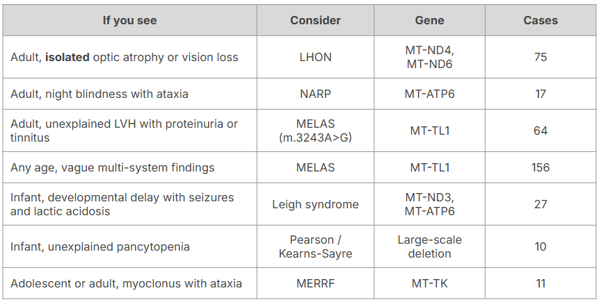
81% of the 348 diagnoses involved just five genes — MT-TL1, MT-ND4, MT-ATP6, MT-ND6, MT-TS1. mtDNA disease is not widely scattered.
5. Why this was possible: mtDNA is in the capture kit
One question remains. How were mtDNA variants found in patients nobody suspected of having mitochondrial disease?
296 of 348 (85%) were diagnosed by exome-based testing; 42 (12%) by genome. No separate mtDNA panel was ordered.
3billion built the full mitochondrial genome into its proprietary exome capture kit. A single exome test reads and interprets nuclear genes and mtDNA together.
Three things follow from that design.
Suspicion isn’t required. The scope of testing isn’t bound to the condition suspected at the time of referral. That is why MELAS surfaced in patients referred for Fabry disease. With a separate panel, you have to suspect it to test for it — and a diagnosis nobody suspected never arrives.
Large-scale deletions are detected. Beyond SNVs, 10 cases of single large-scale mtDNA deletion — the cause of Pearson and Kearns-Sayre syndromes — were identified by the same test.
Both genomes are read at once. In 35 patients (10%), an mtDNA variant was found alongside a nuclear finding: MT-TL1 with TTN, MT-TL1 with GLA, MT-TK with PMP22. Run sequentially, testing stops at whichever finding comes first.
The testing wasn’t split, so the diagnosis wasn’t either.
FAQ
Is exome testing alone sufficient, without a separate mtDNA panel? Of the 348 confirmed patients, 296 (85%) were diagnosed by exome-based testing. 3billion’s exome capture kit includes the full mitochondrial genome, so mtDNA is analyzed without an additional order.
Are single large-scale mtDNA deletions detected? Yes. Ten cases — including Pearson and Kearns-Sayre syndromes — were identified through the same test.
Can a nuclear and a mitochondrial disorder coexist? In 35 patients (10%), both were found. Sequential testing risks stopping at the first finding and missing the second.
Which sample type is appropriate? Standard sample types are suitable, including buccal swab, blood, and DBS. For m.3243A>G specifically, heteroplasmy can be lower in blood, so buccal swab is often considered in adult patients. Contact us for case-specific guidance.
Does this tell us the diagnostic yield or prevalence of mitochondrial disease? No. This is a composition analysis of 348 confirmed patients only. It does not use total tests performed as a denominator, and it should not be read as a yield or prevalence figure.
In closing
This analysis does not tell you how common mitochondrial disease is. It looks only at patients already confirmed, so no yield or prevalence can be calculated — and none is claimed.
What it does show is clear:
- 82% were confirmed without classic mitochondrial findings.
- 75 patients were diagnosed on optic atrophy alone.
- 64 patients referred for suspected Fabry disease had mitochondrial disease.
- Most were identified by a single exome test, with no separate mtDNA panel.
Mitochondrial disease doesn’t arrive looking typical. When testing stops requiring it to, the diagnosis arrives more often.
Based on de-identified aggregate data from 348 cases confirmed at 3billion. No individually identifiable patient information is included.