


Single-Cell Sequencing: Concept, Role, and Trends
Single cell sequencing is a set of high-throughput techniques that measure genomic, transcriptomic, or epigenomic information from individual cells rather than from a pooled tissue sample. This resolution lets researchers separate cell types, spot rare subpopulations, and describe biological heterogeneity that traditional methods obscure.
For clinicians, the practical takeaway is a shift in what a sequencing result can represent. Instead of one averaged profile per sample, single cell approaches return thousands of individual profiles, each tied to one cell.
Frequently asked questions
How does it differ from bulk sequencing?
Bulk sequencing measures the average signal across all cells in a sample. Single cell sequencing measures each cell separately, so it can detect rare subpopulations and heterogeneity that averaging would otherwise hide.
Is single cell sequencing used for rare disease diagnosis?
Not routinely. Today it mainly supports research and mechanism discovery. Standard rare disease diagnosis still relies on exome or genome sequencing. Discuss appropriate testing with a clinician or genetic counselor.
What is single-cell RNA sequencing?
Single-cell RNA sequencing (scRNA-seq) measures gene expression in individual cells. It classifies cell subpopulations and reveals cell heterogeneity, making it the most widely applied form of single cell sequencing.
Can single cell sequencing detect rare cell types?
Yes. Detecting rare or low-abundance cell subpopulations is one of its main strengths, because signals from a few unusual cells are not diluted by the majority population as they are in bulk methods.
Why single cell resolution matters
Tissues are mixtures. A biopsy contains many cell types, states, and lineages, and a disease process may live in only a small fraction of them. Bulk methods report the average across that mixture.
That averaging is the core limitation. Bulk sequencing yields an averaged view of genomic and transcriptomic profiles that can mask rare or low-abundance subpopulations, leaving an incomplete picture of heterogeneity. Single cell methods restore that lost detail.
How it works, at a conceptual level
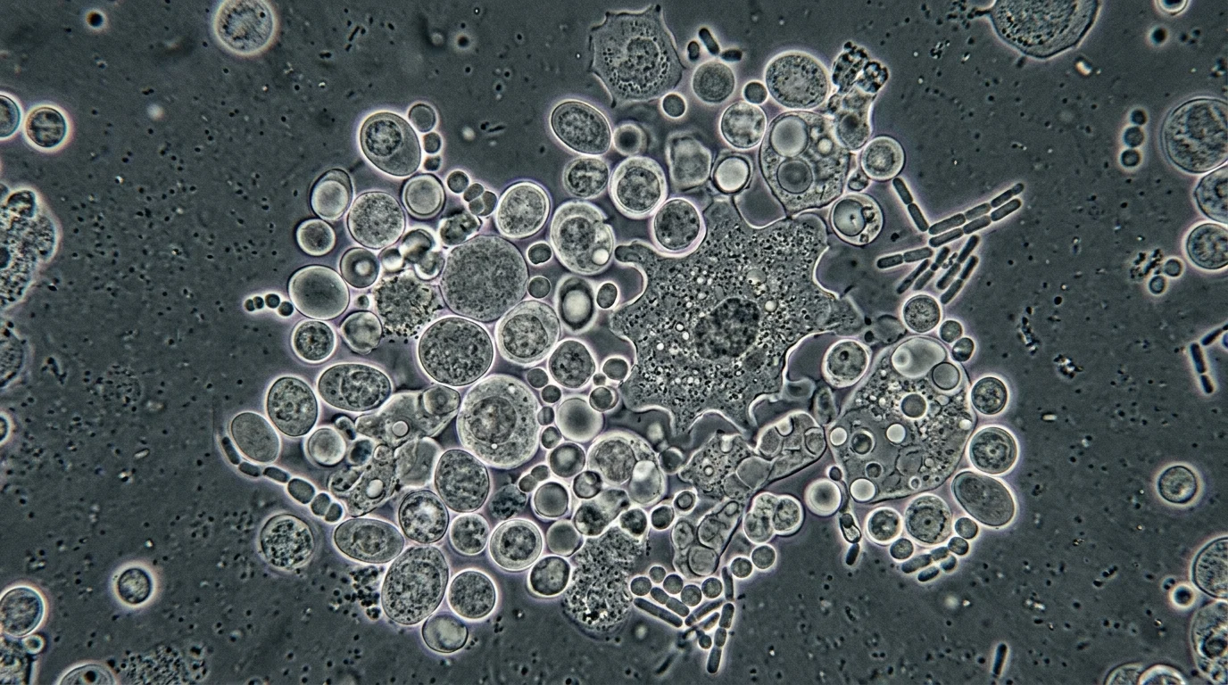
The workflow begins by dissociating a tissue into a suspension of single cells or nuclei. Each cell is then isolated — often in a droplet or well — and its nucleic acids are tagged with a unique molecular barcode before sequencing.
Barcoding is what makes the approach scalable. Because every read carries a cell-of-origin label, thousands of cells can be sequenced together and computationally separated afterward.
What can be measured
Single-cell sequencing detects genomic, transcriptomic, and epigenomic information at the individual-cell level, giving it particular advantage in detecting cellular heterogeneity, classifying cell types precisely, and identifying rare subpopulations. Which layer you measure depends on the biological question.
- Transcriptome — which genes each cell is expressing, and at what level.
- Genome — DNA sequence and copy-number variation cell by cell.
- Epigenome — chromatin accessibility or methylation states per cell.
Single-cell RNA sequencing in practice
The transcriptomic version, scRNA-seq, is the most mature and widely used. scRNA-seq has become a powerful tool for cell subpopulation classification and describing cell heterogeneity through high-throughput, multidimensional analysis of individual cells, circumventing the averaging limitation of traditional sequencing.
In practice, this lets investigators build cell atlases of a tissue, trace developmental trajectories, and pinpoint the specific cell populations perturbed in a disease state. For rare disease research, that can clarify how a variant in a broadly expressed gene produces a tissue-restricted phenotype.

Toward single-cell multi-omics
The field no longer stops at RNA. Single-cell sequencing has expanded to include epigenomics, proteomics, metabolomics, and microbiome analysis, and integrating these layers has advanced single-cell multi-omics sequencing.
Measuring several molecular layers in the same cell links genotype, regulatory state, and functional output more directly. That integration is where much of the current methodological momentum sits.
Where it fits in rare disease work today
It helps to separate diagnostics from discovery. Clinical diagnosis of a suspected monogenic rare disease still rests on exome or genome sequencing, which interrogate the germline variant present in every nucleated cell.
Single cell sequencing instead earns its place in mechanism research: functional follow-up of variants of uncertain significance, characterizing affected tissues, and understanding why a given genotype produces a specific cellular phenotype. Think of it as a tool that explains disease biology rather than one that returns a routine diagnostic report.
References
- Chen S, et al., Application of single-cell sequencing to the research of tumor microenvironment, 2023, DOI: 10.3389/fimmu.2023.1285540, https://pmc.ncbi.nlm.nih.gov/articles/PMC10641410/
- Wang S, et al., The Evolution of Single-Cell RNA Sequencing Technology and Application: Progress and Perspectives, 2023, DOI: 10.3390/ijms24032943, https://pmc.ncbi.nlm.nih.gov/articles/PMC9918030/
- Zhang K, et al., Advancement of single-cell sequencing for clinical diagnosis and treatment of pancreatic cancer, 2023, DOI: 10.3389/fmed.2023.1213136, https://pmc.ncbi.nlm.nih.gov/articles/PMC10501729/
- Zhang W, et al., Research progress and the prospect of using single-cell sequencing technology to explore the characteristics of the tumor microenvironment, 2024, DOI: 10.1016/j.gendis.2024.101239, https://pmc.ncbi.nlm.nih.gov/articles/PMC11566696/

Soo-jung Baek
Marketing Manager
I strive to empower the rare disease community by sharing meaningful insights backed by our company’s expertise.