



Cloverleaf Skull: Is It Pfeiffer Syndrome Type 2?
*This article has been reviewed by a clinical geneticist at 3billion.
Pfeiffer syndrome type 2 is the most severe subtype of a rare craniosynostosis disorder, distinguished by a trilobed cloverleaf skull, extreme proptosis, and broad thumbs and great toes. It arises from de novo heterozygous mutations in the FGFR2 gene and typically presents with developmental delay, neurological complications, and life-threatening airway compromise.
For clinicians, the practical challenge is twofold: recognizing the phenotype early—often prenatally—and confirming the molecular diagnosis to separate it from phenotypic mimics such as thanatophoric dysplasia. Pfeiffer syndrome as a whole affects roughly 1 in 100,000 individuals, and type 2 represents a small fraction of that population.
Frequently asked questions
How does type 2 differ from type 3?
Types 2 and 3 share severe features, but only type 2 shows a cloverleaf skull caused by more extensive premature fusion of skull bones. Type 3 presents similar systemic findings without this trilobed cranial deformity.
Which gene causes Pfeiffer syndrome type 2?
Pfeiffer syndrome type 2 is caused almost exclusively by de novo heterozygous mutations in the FGFR2 gene.
Is Pfeiffer syndrome type 2 inherited?
Type 2 nearly always arises from new (de novo) sporadic mutations rather than inheritance from a parent. Advanced paternal age raises risk because of an increased likelihood of new FGFR mutations in paternal sperm.
What is the prognosis for Pfeiffer syndrome type 2?
Prognosis is guarded. Unlike the milder type 1, type 2 often causes significant neurological impairment and early mortality, largely driven by airway compromise and complications of severe craniosynostosis.
The Pfeiffer spectrum: where type 2 sits
Pfeiffer syndrome is classically divided into three clinical subtypes that share the core triad of craniosynostosis, broad thumbs, and broad great toes but diverge sharply in severity.
Type 1, the classic form, is associated with normal intelligence and a generally favorable outcome. Types 2 and 3 are far more severe, often leading to significant neurological impairment and early mortality.
The dividing line between the two severe subtypes is anatomical. Type 2 consists of cloverleaf skull, extreme proptosis, finger and toe abnormalities, elbow ankylosis or synostosis, developmental delay, and neurological complications; type 3 is similar to type 2 but without a cloverleaf skull.
Put simply, the presence of the cloverleaf deformity is what separates type 2 from type 3. That distinction reflects more extensive fusion of bones in the skull.
Molecular basis: FGFR2 and the FGF signaling axis
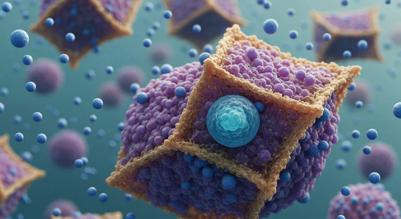
The fibroblast growth factor receptors are transmembrane tyrosine kinases that regulate osteoblast proliferation and cranial suture patterning. Gain-of-function mutations produce constitutive or ligand-independent receptor activation, driving premature ossification of the sutures.
Pfeiffer syndrome is most commonly caused by mutations in the FGFR2 gene, while FGFR1 mutations account for only a small percentage of type 1 cases and have not been associated with type 2 or 3. For the severe subtypes, FGFR2 is effectively the relevant locus.
Type 2 is caused almost exclusively by de novo heterozygous mutations in the FGFR2 gene. No single variant accounts for most cases, but a limited number of recurrent pathogenic variants—W290C, Y340C, C342R, and S351C—are associated with the severe subtypes. Reported pathogenic variants include missense changes such as c.335A>G (p.Tyr112Cys) and c.1019A>G (p.Y340C), the latter documented in an autopsy case with cloverleaf skull, hydrocephalus, and a tracheal cartilage sleeve.
Because these variants arise de novo, advanced paternal age is a recognized risk factor owing to the increased likelihood of new FGFR mutations. Recurrence risk for future pregnancies of unaffected parents is low but not zero, given the possibility of parental germline mosaicism.
Clinical recognition
Cranial and craniofacial features
The cloverleaf skull—a trilobed cranium from fusion of multiple sutures—is the defining sign of type 2. It results from premature fusion of all sutures and has been detected on prenatal ultrasound as early as 21–22 weeks gestation. Extreme proptosis follows from shallow orbits and can threaten corneal integrity and vision.
Limb and skeletal findings
Broad, medially deviated thumbs and great toes are characteristic. Elbow ankylosis or radiohumeral synostosis restricts joint mobility and is a useful discriminating feature at examination.
Neurological and airway complications
Hydrocephalus, Chiari malformation, and raised intracranial pressure contribute to developmental delay and neurological morbidity. Upper airway anomalies are a leading cause of death.
Diagnostic workup
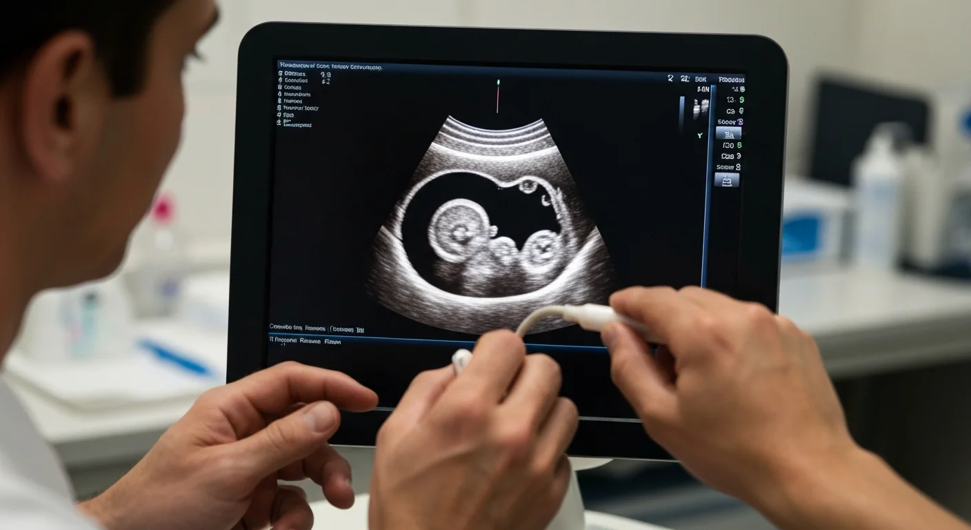
Suspicion often begins prenatally. Fetuses carrying a Y340C FGFR2 mutation may present a cloverleaf skull on prenatal ultrasound, and whole exome sequencing is useful for rapid differential diagnosis of Pfeiffer syndrome from thanatophoric dysplasia type II. That distinction changes counseling and delivery planning.
A pragmatic diagnostic sequence for clinicians:
- Detailed fetal or neonatal imaging to characterize suture involvement and orbital depth.
- Targeted FGFR2 analysis or a craniosynostosis gene panel when the phenotype is classic.
- Rapid whole exome or genome sequencing when the differential is broad or a fast answer is needed for perinatal management.
- Variant classification against ACMG criteria; several Pfeiffer-associated variants have been reclassified to definitively pathogenic on review.
Molecular confirmation does more than label the condition. It clarifies subtype, supports recurrence-risk counseling, and can retrospectively refine an antenatal impression once postnatal testing is complete.
When to refer and counsel
Any neonate or fetus with multisuture craniosynostosis, cloverleaf skull, or proptosis with broad thumbs warrants prompt referral to clinical genetics and a craniofacial team. Multidisciplinary input—neurosurgery, ophthalmology, and airway specialists—should be mobilized early given the risk profile.
If you need an accurate diagnosis through whole exome sequencing, 3billion is your best choice. We provide automated reanalysis at no additional cost and with no separate request required. Click the button below to send us your questions about test pricing, the process, and anything else. We’ll get back to you promptly.
Want to learn more about
3billion's genetic testing?
We'll reply within 1 business day.
This article is not diagnostic advice. Actual testing and diagnosis should be decided together with a clinician or genetic counselor.
References
- Vogels A, Fryns JP, “Pfeiffer syndrome”, Orphanet J Rare Dis. 2006;1:19, DOI: 10.1186/1750-1172-1-19, https://pmc.ncbi.nlm.nih.gov/articles/PMC1482682/
- Mosalli R, Fatma A, Almatrafi MA, Mazroua M, Paes B, “De Novo Heterozygous Mutation in FGFR2 Causing Type II Pfeiffer Syndrome”, Case Rep Genet. 2022;2022:4791082, DOI: 10.1155/2022/4791082, https://pmc.ncbi.nlm.nih.gov/articles/PMC9537020/
- Katsuragi SY, Hirose E, Arai Y, Otsuki Y, Ohki S, Kobayashi H, “Autopsy Case of Pfeiffer Syndrome Type 2, a Phenotype of Fibroblast Growth Factor Receptor-Associated Craniosynostosis Syndromes, with Tracheal Cartilage Sleeve and Abnormal Hyperplasia of Bronchial Cartilages”, Am J Case Rep. 2021;22:e932450, DOI: 10.12659/AJCR.932450, https://pmc.ncbi.nlm.nih.gov/articles/PMC8363655/
- Chen CP, Huang JP, Huang KS, Chen YY, Wu FT, Pan YT, Chiu CL, Wang W, “Perinatal imaging findings of a fetus with Pfeiffer syndrome and a heterozygous c.1019A>G, p.Tyr340Cys (Y340C) mutation in FGFR2 presenting a cloverleaf skull, craniosynostosis and short limbs on prenatal ultrasound mimicking thanatophoric dysplasia type II”, Taiwan J Obstet Gynecol. 2024;63(3):387–390, DOI: 10.1016/j.tjog.2024.03.005, https://pubmed.ncbi.nlm.nih.gov/38802203/
- Oyamada MK, Ferreira HSA, Hoff M, “Pfeiffer syndrome type 2 – case report”, São Paulo Med J. 2003;121(4), DOI: 10.1590/S1516-31802003000400008, https://www.ncbi.nlm.nih.gov/pmc/articles/PMC11108598/

Soo-jung Baek
Marketing Manager
I strive to empower the rare disease community by sharing meaningful insights backed by our company’s expertise.