



Hypochondroplasia vs. Achondroplasia: What Distinguishes Them

Sookjin Lee
Chief Business Officer (CBO) | Ph.D.
A business expert leading innovation in precision medicine and genomics for 20 years. Accelerating the commercialization of innovative technologies with academic expertise and market insight.
Key summary
- Same gene, different position. Achondroplasia arises at FGFR3 c.1138 (p.Gly380Arg); hypochondroplasia most often at c.1620 (p.Asn540Lys). A c.1138-only assay cannot detect the latter.
- Testing both positions is what guidance recommends — and it still leaves a gap. Targeted analysis of the c.1620 pair identifies ~70–80% of cases; full FGFR3 sequencing 70–90%; in 10–30% no FGFR3 variant is found.
- Different surveillance, not just milder. Foramen magnum stenosis, spinal stenosis, tibial bowing and obstructive apnea occur less often than in achondroplasia. Temporal lobe epilepsy is the concern that rises.
1. Two conditions, one gene, two hotspots
Achondroplasia has an unusually concentrated molecular basis. More than 99% of patients carry one of two changes at a single position — c.1138G>A or c.1138G>C — and both produce p.Gly380Arg [9]. That concentration is what makes targeted testing so efficient, and what makes it a trap in the condition next to it.
The genetics, diagnosis and treatment of achondroplasia itself are covered in Achondroplasia in children: from the FGFR3 mutation to Voxzogo.
Hypochondroplasia is also caused by FGFR3, but the recurrent variants sit elsewhere: c.1620C>A and c.1620C>G, both encoding p.Asn540Lys (N540K in the literature) [1]. The architecture is a mirror image — two substitutions at one nucleotide producing one amino acid change — but the nucleotide is a different one.
So a c.1138 assay comes back negative in a patient who has hypochondroplasia. The result is not wrong. It answered the question it was asked. It did not answer the question the clinician had.
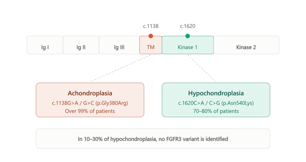
GeneReviews is direct about the consequence. Because the two conditions may present very similarly and are easily confused, both pairs of variants should be tested when molecular testing is requested for hypochondroplasia [1].
One further point about this gene: the p.Lys650 codon appears in thanatophoric dysplasia, in SADDAN, and among hypochondroplasia variants [1]. A single codon spans a lethal condition and one of the mildest in the series. Position does not predict phenotype here.
2. A case in which hotspot testing delayed the diagnosis
In a published case report, a fetus showed shortened limbs below the 3rd percentile, an increased biparietal diameter above the 95th percentile, and a low nasal bridge on serial ultrasound. Given the relative frequency of achondroplasia, targeted testing for the FGFR3 c.1138 hotspot was performed in the third trimester. It was negative [2].
The child continued to show growth retardation, rhizomelic disproportionate short stature, an enlarged head and short hands. Whole exome sequencing at one year of age identified FGFR3 c.1620C>A, and the diagnosis was hypochondroplasia [2]. The authors’ conclusion is worth stating plainly: hotspot testing should not serve as the sole basis for a clinical diagnosis, and where clinical findings conflict with a negative targeted result, further analysis should follow promptly rather than be deferred [2].
Current guidance anticipates this. Hypochondroplasia is typically suspected only late in the third trimester, from short long bones and a larger head circumference. Where those findings are present, prenatal imaging may not demonstrate all features, and a broad multigene panel including FGFR3, or comprehensive genomic testing, can be considered on a fetal sample [1,3].
A normal third-trimester ultrasound does not exclude the diagnosis [1]. That is an argument against stopping once there was a reason to suspect — not an argument for testing pregnancies in which no suspicious finding was seen.
3. Testing both hotspots helps — and still leaves a gap
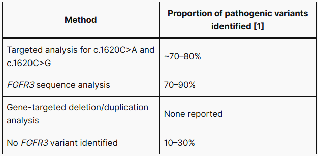
Two implications follow.
Full-gene sequencing outperforms hotspot analysis. Pathogenic variants causing hypochondroplasia also occur outside c.1620: c.829A>G (p.Tyr278Cys) resembles achondroplasia in the newborn period, and c.1043C>G (p.Ser348Cys) is severe enough to be described as a mild-achondroplasia phenotype [1]. A c.1620-only assay will not detect them.
For 10–30% of patients the gene is not the answer. Locus heterogeneity is considered possible, and the diagnostic criteria themselves remain contested — no clinical or radiologic feature is unique to hypochondroplasia, and there is no consensus on how many are required [1].
There is a second problem behind the first. Patients with p.Asn540Lys tend to have more pronounced findings than those without it [1]. Patients whose hotspot test comes back negative are therefore often the milder cases — a negative result and an ambiguous phenotype together, which is where follow-up tends to stop.
4. What differs clinically
Hypochondroplasia is often called a milder achondroplasia. That is accurate about stature. It does not follow that there is less to watch for.
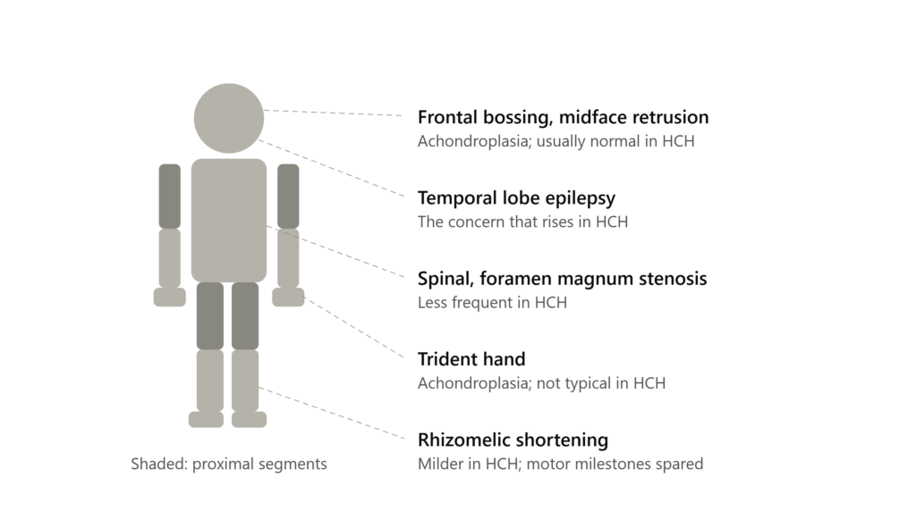
Face and hands — hypochondroplasia has no distinguishing features. Craniofacial appearance is usually normal. The classic achondroplasia features, midface retrusion and frontal bossing, are not generally seen. The hands are relatively short but do not show the trident configuration [1]. Short stature without these features should raise hypochondroplasia as a possibility.
Skeletal complications — less frequent in hypochondroplasia. Foramen magnum stenosis, spinal stenosis, tibial bowing and obstructive apnea are all less common than in achondroplasia. Spinal stenosis symptoms in adults are milder, and motor milestones are usually not significantly delayed [1].
Epilepsy — more to watch for in hypochondroplasia. Seizures can present at any age and typically have a temporal lobe focus. In infants the presentation is easy to miss: apneas, cyanosis, eye deviation, vacant stares, with EEG findings that can be difficult to capture [1]. Temporal lobe dysgenesis has been reported in FGFR3-related conditions, including incomplete hippocampal folding and dilatation of the temporal horns [1].
How infant apnea is interpreted matters here. In achondroplasia it is commonly airway obstruction or foramen magnum stenosis. In hypochondroplasia it may be a temporal lobe seizure.
Intelligence — not settled. Intellectual disability is thought to be more common in hypochondroplasia, but GeneReviews notes that robust data are scarce and the observation has been controversial [1]. Two cohorts are cited. Among 13 Finnish individuals with confirmed p.Asn540Lys, eight had neurocognitive difficulties: specific learning disorder in two, mild intellectual disability in five, global developmental delay in one [4]. A cohort of 20 molecularly confirmed Korean patients reported developmental delay in 25% [5]. The numbers are too small to generalize from.
In short. Neither condition is simply worse than the other. They have different things to monitor. A patient carried as “mild achondroplasia” is checked repeatedly for complications they are unlikely to develop, while temporal lobe epilepsy never enters the list.
5. Which conditions to rule out
The differential matters more on the hypochondroplasia side. At its mildest the phenotype is indistinguishable from idiopathic or familial short stature [1], and children typically present to pediatrics or pediatric endocrinology as toddlers or at early school age with nothing more specific than decreased growth velocity. Achondroplasia, by contrast, is 99% one position — a single targeted test usually settles it.
Conditions with a known genetic etiology that may be confused with hypochondroplasia [1]:
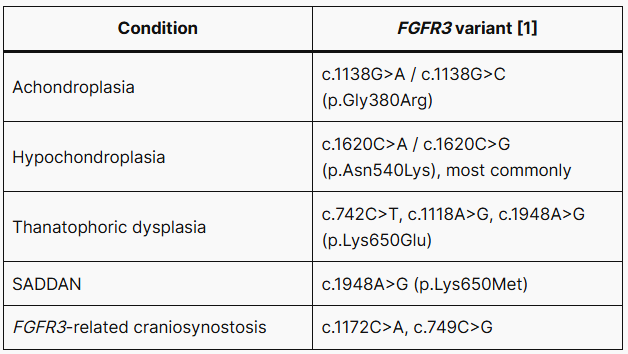
Short stature from disturbances of the growth hormone axis and constitutional short stature are also considered [1]. Where the presentation is more achondroplasia-like, the list changes: pseudoachondroplasia (COMP ) and spondyloepiphyseal dysplasia congenita (COL2A1 ) enter instead.
These conditions have different causal genes and several are recessive. A single-gene assay cannot arbitrate between them.
6. Why the distinction is becoming therapeutically consequential
Until recently, precise subtyping changed surveillance but not therapy. That is changing.
- Vosoritide in hypochondroplasia. A phase 2 trial in children has been published [6]. A phase 3 trial met its primary endpoint in May 2026, with annualized growth velocity improved by +2.33 cm/yr, and a supplemental FDA application was planned for Q3 2026. Hypochondroplasia is not an approved indication at the time of writing.
- Oral FGFR3 inhibition. Infigratinib met its annualized height velocity endpoint in phase 3 in achondroplasia, with submissions planned for H2 2026 and stated plans to accelerate development in hypochondroplasia.
- Longer-acting CNP analogues, including navepegritide, which has reported phase 2 results in achondroplasia. Not every program has succeeded — the FGFR3 ligand trap recifercept was discontinued after an interim analysis suggested futility.
For achondroplasia itself, consensus guidelines on implementing and monitoring vosoritide were published in 2025 [8].
The implication is concrete: eligibility for these trials, and in time for approved therapy, is defined molecularly rather than radiographically. A patient carried as “probable achondroplasia, hotspot-negative” is on neither pathway.molecular result does not confirm the clinical label — it determines which label applies.
7. What the guidance supports
The International Consensus Statement on achondroplasia notes that genetic testing helps distinguish achondroplasia from hypochondroplasia and other skeletal dysplasias, and that skeletal dysplasia gene panels or whole exome sequencing can be used in patients with atypical clinical and/or radiographic presentations [7]. GeneReviews sets out the same division for hypochondroplasia: gene-targeted testing where the phenotype and radiographs suggest the diagnosis, and comprehensive genomic testing where the phenotype is indistinguishable from many other skeletal dysplasias [1].
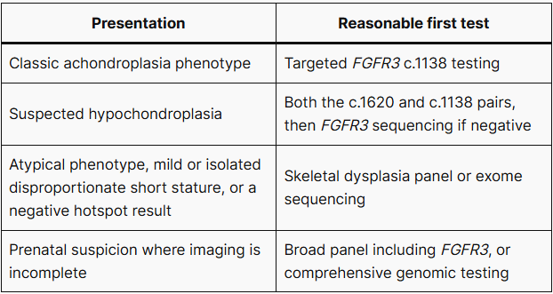
Exome sequencing is not the last step. Where the phenotype remains convincing and coding analysis is uninformative, genome sequencing covers the deep intronic and regulatory regions that exome capture cannot reach — one case report describes clinical and radiographic achondroplasia resolved only by extending FGFR3 analysis into non-coding sequence [9]. Reanalysis as databases evolve is worthwhile in unsolved cases, and DNA banking is recommended where no molecular diagnosis has been established [1].
Frequently asked questions
Does a negative FGFR3 c.1138 test exclude achondroplasia? It makes classic achondroplasia very unlikely, since more than 99% of patients carry a variant at that position [9]. It does not exclude hypochondroplasia, another FGFR3-related condition, or a skeletal dysplasia caused by a different gene. If the phenotype was convincing enough to prompt testing, a negative result is a reason to broaden rather than to stop.
If I add the c.1620 hotspot, is that enough? Testing both positions is what guidance recommends, precisely because the two conditions are easily confused [1]. It is not complete: targeted analysis of the c.1620 pair identifies roughly 70–80% of hypochondroplasia, full FGFR3 sequencing 70–90%, and in 10–30% of patients no FGFR3 variant is identified [1].
When the hotspot is negative: 3B-EXOME
Whole exome sequencing with variant interpretation across the full skeletal dysplasia gene set, for patients whose presentation does not fit a single-gene assay.
References
- Bober MB, Bellus GA, Cheung MS, Jain M, Nikkel SM, Tiller GE. Hypochondroplasia. In: Adam MP, Bick S, Mirzaa GM, et al, eds. GeneReviews. Seattle, WA: University of Washington, Seattle; 1993-2026. Updated September 25, 2025. Accessed July 27, 2026. https://www.ncbi.nlm.nih.gov/books/NBK1477/
- Xie H, Chen Y, Xiong F, Li J, Yang F. Failure to diagnose hypochondroplasia by prenatal diagnosis: a case report. BMC Pediatr. 2023;23(1):100. doi:10.1186/s12887-023-03917-2
- Sabir AH, Sheikh J, Singh A, et al. Earlier detection of hypochondroplasia: a large single-center UK case series and systematic review. Am J Med Genet A. 2021;185(1):73-82. doi:10.1002/ajmg.a.61912
- Linnankivi T, Mäkitie O, Valanne L, Toiviainen-Salo S. Neuroimaging and neurological findings in patients with hypochondroplasia and FGFR3 N540K mutation. Am J Med Genet A. 2012;158A:3119-3125. PMID:23165795
- Kim HY, Lee YA, Shin CH, Cho TJ, Ko JM. Clinical manifestations and outcomes of 20 Korean hypochondroplasia patients with the FGFR3 N540K variant. Exp Clin Endocrinol Diabetes. 2023;131:123-131. PMID:36442838
- Dauber A, Zhang A, Kanakatti Shankar R, et al. Vosoritide treatment for children with hypochondroplasia: a phase 2 trial. EClinicalMedicine. 2024;71:102591. PMID:38813446
- Savarirayan R, Ireland P, Irving M, et al. International consensus statement on the diagnosis, multidisciplinary management and lifelong care of individuals with achondroplasia. Nat Rev Endocrinol. 2022;18(3):173-189. doi:10.1038/s41574-021-00595-x
- Savarirayan R, Hoover-Fong J, Ozono K, et al. International consensus guidelines on the implementation and monitoring of vosoritide therapy in individuals with achondroplasia. Nat Rev Endocrinol. 2025;21(5):314-324. doi:10.1038/s41574-024-01074-9
- Ouedraogo ZG, Janel C, Janin A, et al. Relevance of extending FGFR3 gene analysis in osteochondrodysplasia to non-coding sequences: a case report. Genes (Basel). 2024;15(2):225. doi:10.3390/genes15020225